

Characterization of T cell repertoire of blood, tumor, and ascites in ovarian cancer patients using next generation sequencing
- PMID: 26451311
- PMCID: PMC4589054
- DOI: 10.1080/2162402X.2015.1030561
Characterization of T cell repertoire of blood, tumor, and ascites in ovarian cancer patients using next generation sequencing
Abstract
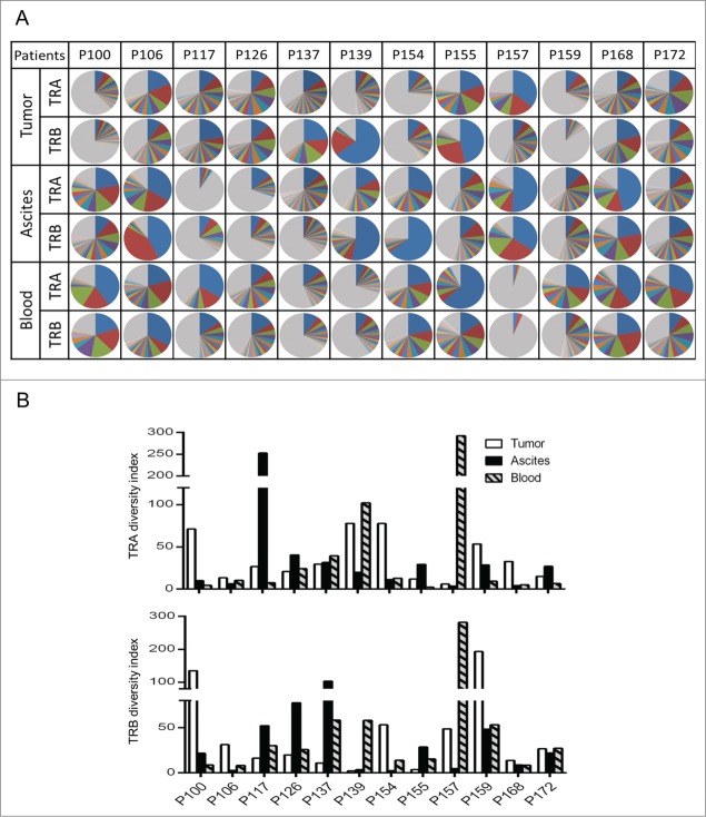
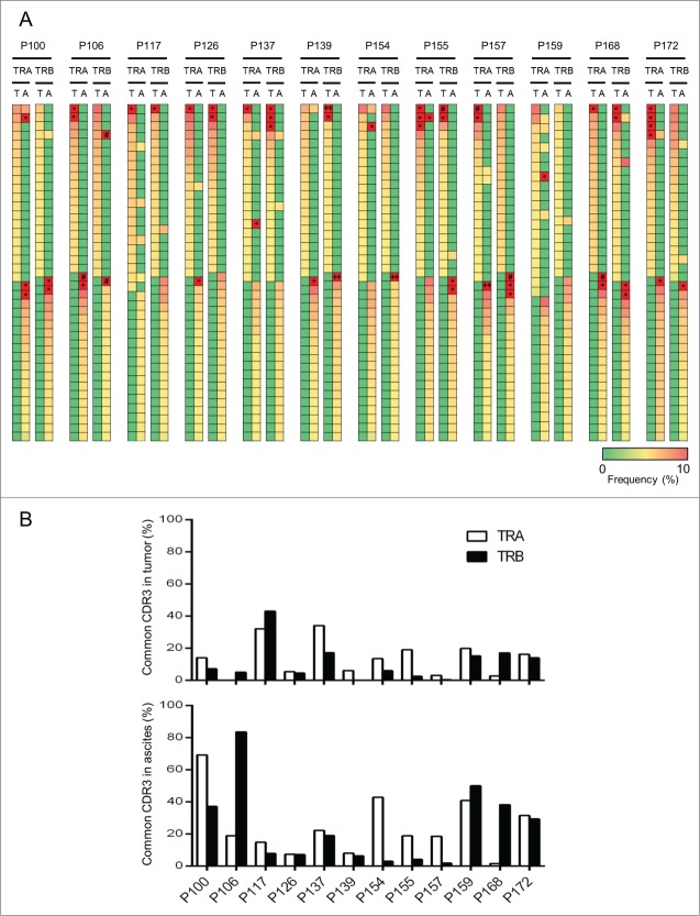
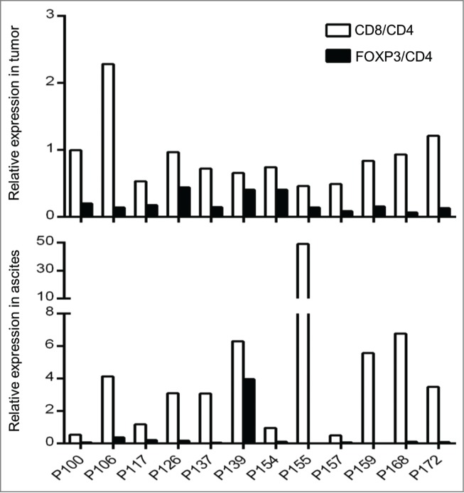
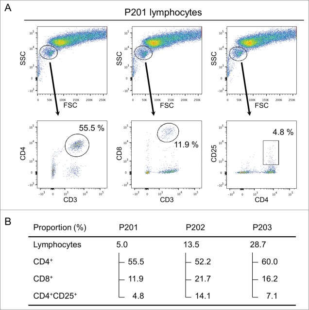
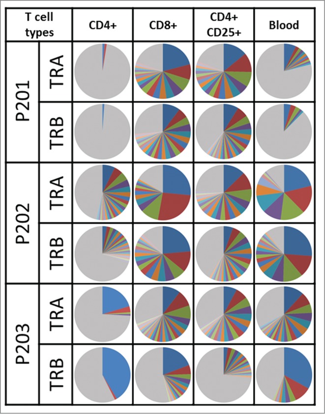
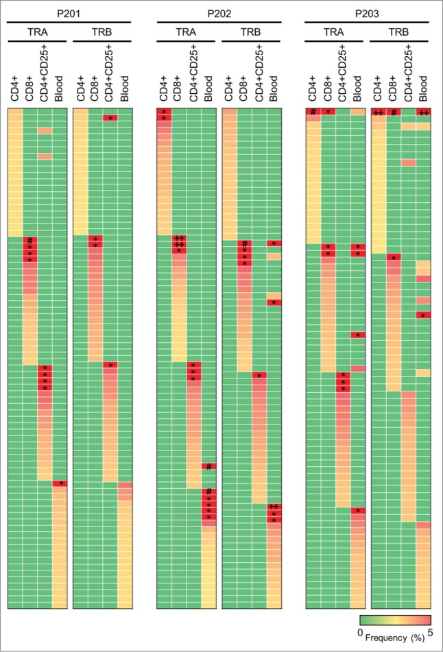
Tumor-infiltrating lymphocytes (TILs) play an important role in regulating the host immune response and are one of key factors in defining tumor microenvironment. Some studies have indicated that T cell infiltration in malignant ascites is associated with clinical outcome, but few studies have performed detailed characterization of T cell diversity or clonality in malignant effusions. We have applied a next generation sequencing method to characterize T cell repertoire of a set of primary cancers, ascites, and blood from 12 ovarian cancer patients and also analyzed the T cell subtype populations in malignant fluids from 3 ovarian cancer patients. We observed enrichment of certain T cells in tumors and ascites, but most of the enriched T cell receptor (TCR) sequences in tumors and ascites were not common. Moreover, we analyzed TCR sequences of T cell subtypes (CD4+, CD8+, and regulatory T cells) isolated from malignant effusions and also found clonal expansion of certain T cell populations, but the TCR sequences were almost mutually exclusive among the three subgroups. Although functional studies of clonally expanded T cell populations are definitely required, our approach offers a detailed characterization of T cell immune microenvironment in tumors and ascites that might differently affect antitumor immune response.
Keywords: T cell receptor; malignant ascites; next generation sequencing; ovarian cancer; tumor immunity.
Figures
References
-
- Siegel R, Ma J, Zou Z, Jemal A. Cancer statistics, 2014. CA Cancer J Clin 2014; 64:9-29; PMID:24399786; http://dx.doi.org/10.3322/caac.21208 - DOI - PubMed
-
- Ayhan A, Gultekin M, Taskiran C, Dursun P, Firat P, Bozdag G, Celik NY, Yuce K. Ascites and epithelial ovarian cancers: a reappraisal with respect to different aspects. Int J Gynecol Cancer 2007; 17:68-75; PMID:17291234; http://dx.doi.org/10.1111/j.1525-1438.2006.00777.x - DOI - PubMed
-
- Parsons SL, Lang MW, Steele RJ. Malignant ascites: a 2-year review from a teaching hospital. Eur J Surg Oncol 1996; 22:237-9; PMID:8654603; http://dx.doi.org/10.1016/S0748-7983(96)80009-6 - DOI - PubMed
-
- Sheid B. Angiogenic effects of macrophages isolated from ascitic fluid aspirated from women with advanced ovarian cancer. Cancer Lett 1992; 62:153-8; PMID:1371714; http://dx.doi.org/10.1016/0304-3835(92)90186-Y - DOI - PubMed
-
- Ioannides CG, Platsoucas CD, Rashed S, Wharton JT, Edwards CL, Freedman RS. Tumor cytolysis by lymphocytes infiltrating ovarian malignant ascites. Cancer Res 1991; 51:4257-65; PMID:1868446 - PubMed
Publication types
LinkOut - more resources
Full Text Sources
Other Literature Sources
Molecular Biology Databases
Research Materials