

Diagnosis and Management of Iridocorneal Endothelial Syndrome
- PMID: 26451377
- PMCID: PMC4588350
- DOI: 10.1155/2015/763093
Diagnosis and Management of Iridocorneal Endothelial Syndrome
Abstract
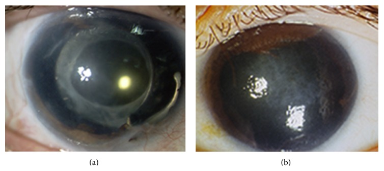
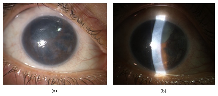
The iridocorneal endothelial (ICE) syndrome is a rare ocular disorder that includes a group of conditions characterized by structural and proliferative abnormalities of the corneal endothelium, the anterior chamber angle, and the iris. Common clinical features include corneal edema, secondary glaucoma, iris atrophy, and pupillary anomalies, ranging from distortion to polycoria. The main subtypes of this syndrome are the progressive iris atrophy, the Cogan-Reese syndrome, and the Chandler syndrome. ICE syndrome is usually diagnosed in women in the adult age. Clinical history and complete eye examination including tonometry and gonioscopy are necessary to reach a diagnosis. Imaging techniques, such as in vivo confocal microscopy and ultrasound biomicroscopy, are used to confirm the diagnosis by revealing the presence of "ICE-cells" on the corneal endothelium and the structural changes of the anterior chamber angle. An early diagnosis is helpful to better manage the most challenging complications such as secondary glaucoma and corneal edema. Treatment of ICE-related glaucoma often requires glaucoma filtering surgery with antifibrotic agents and the use of glaucoma drainage implants should be considered early in the management of these patients. Visual impairment and pain associated with corneal edema can be successfully managed with endothelial keratoplasty.
Figures
References
-
- Harms C. Einseitige spontone Liickenbildung der Iris durch Atrophie ohne mechanische Zerrung. Klinische Monatsblätter für Augenheilkunde. 1903;41:522–528.
-
- Lohelein H. Ztur Kenntnis der essentiellen fortschreitenideni Irisatrophie Illit Lochbildung unld Glaukom. Klin Monbl Augenheilkd Augenarztl Fortbild. 1951;118:379–388. - PubMed
Publication types
MeSH terms
LinkOut - more resources
Full Text Sources
Other Literature Sources
