. 2015 Sep 1;1(2):103-110.
doi: 10.1016/j.trci.2015.07.001.

Marked gender differences in progression of mild cognitive impairment over 8 years

Affiliations

Marked gender differences in progression of mild cognitive impairment over 8 years

Katherine A Lin et al. Alzheimers Dement (N Y). .

Abstract

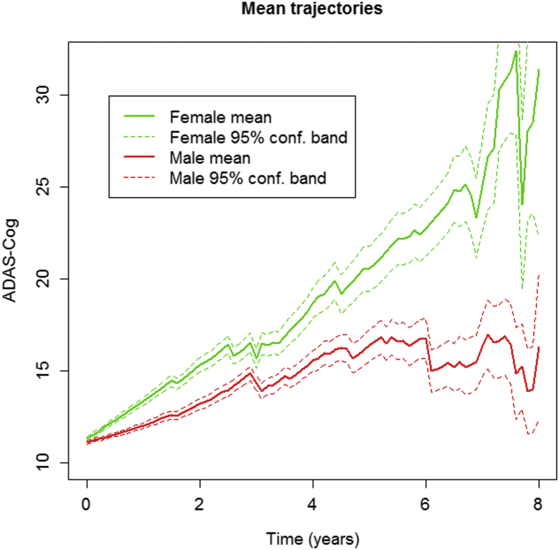
Introduction: This study examined whether, among subjects with mild cognitive impairment (MCI), women progressed at faster rates than men.

Methods: We examine longitudinal rates of change from baseline in 398 MCI subjects (141 Females, 257 Males) in the Alzheimer's Disease Neuroimaging Initiative-1 (ADNI-1), followed for up to 8 years (mean 4.1±2.5 years) using mixed effects models incorporating all follow ups (mean 8±4 visits).

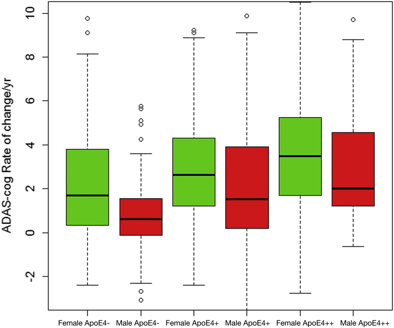
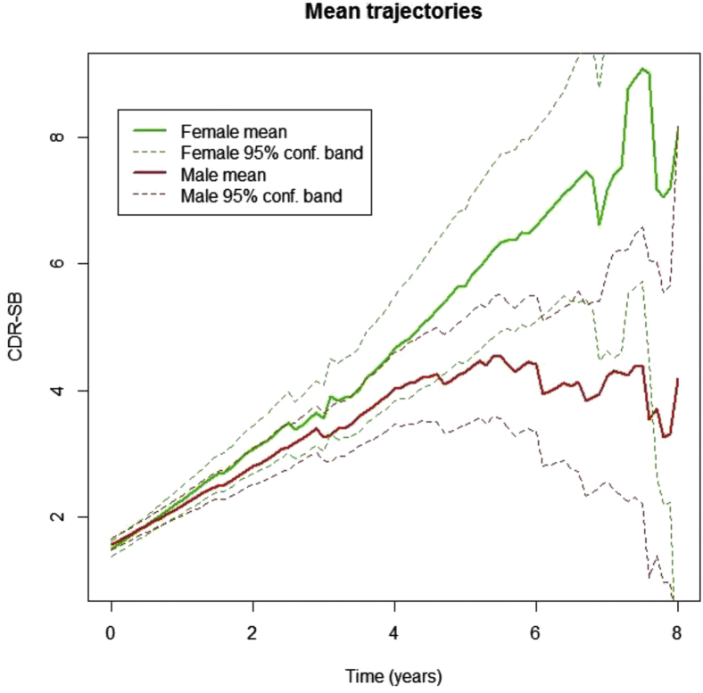
Results: Women progressed at faster rates than men on ADAS-Cog (p=0.001) and CDR-SB (p=0.003). Quadratic fit for change over time was significant for both ADAS-Cog (p=0.001) and CDR-SB (p=0.004), and the additional acceleration in women was 100% for ADAS-Cog and 143% for CDR-SB. The variability of change was greater in women. The gender effect was greater in ApoE4 carriers.

Discussion: Women with MCI have greater longitudinal rates of cognitive and functional progression than men. Studies to confirm and uncover potential mechanisms appear to be warranted.

Keywords: disease modification; gender differences; prevalence; secondary prevention.


Figures

Fig. 1
Fig. 1
ADAS-Cog changes over time by gender. Average trajectories of ADAS-Cog scores by gender. Pointwise 95% confidence bands for the mean are based on the number of subjects at any given time point. The figure depicts time in years from baseline on the x-axis and ADAS-Cog 11 total scores on the y-axis. Increasing ADAS-Cog scores indicate worsening. Solid lines indicate mean ADAS-Cog scores, and dashed lines indicate 95% confidence intervals for these scores. The actual visit date of each subject was used (rather than pooling visits as “annual”) to give a more precise depiction of variability and progression. Abbreviation: ADAS-Cog, Alzheimer's disease assessment scale-cognitive subscale.
Fig. 2
Fig. 2
Median rates of ADAS-Cog change by gender and APOE ε4 status. Median rates of ADAS-Cog change per year by gender and number of APOE ε4 alleles. APOE ε4− indicates no APOE ε4 alleles, APOE ε4+ is one allele, and APOE ε4++ is two alleles. Women had higher median annual rate of change than men regardless of APOE ε4 genotype group. Abbreviation: ADAS-Cog, Alzheimer's disease assessment scale-cognitive subscale.
Fig. 3
Fig. 3
CDR-SB changes over time by gender. Average trajectories of CDR-SB scores by gender. Pointwise 95% confidence bands for the mean are based on the number of subjects at risk at any given time point. The figure depicts time in years from baseline on the x-axis and CDR-SB scores on the y-axis. Higher CDR-SB scores indicate worsening. Solid lines indicate mean ADAS-Cog scores, and dashed lines indicate 95% confidence intervals for these scores. The actual visit date of each subject was used (rather than pooling visits as “annual”) to give a more precise depiction of variability and progression. Abbreviations: CDR-SB, clinical dementia rating-sum of boxes; ADAS-Cog, Alzheimer's disease assessment scale-cognitive subscale.

References

    1. Mielke M.M., Vemuri P., Rocca W.A. Clinical epidemiology of Alzheimer's disease: Assessing sex and gender differences. Clin Epidemiol. 2014;6:37–48. - PMC - PubMed
    1. Hebert L.E., Weuve J., Scherr P.A., Evans D.A. Alzheimer disease in the United States (2010-2050) estimated using the 2010 census. Neurology. 2013;80:1778–1783. - PMC - PubMed
    1. Seshadri S., Wolf P.A., Beiser A., Au R., McNulty K., White R. Lifetime risk of dementia and Alzheimer's disease. The impact of mortality on risk estimates in the Framingham Study. Neurology. 1997;49:1498–1504. - PubMed
    1. Hua X., Hibar D.P., Lee S., Toga A.W., Jack C.R., Jr., Weiner M.W. Sex and age differences in atrophic rates: An ADNI study with n1368 MRI scans. Neurobiol Aging. 2010;31:1463–1480. - PMC - PubMed
    1. Barnes L.L., Wilson R.S., Bienias J.L., Schneider J.A., Evans D.A., Bennett D.A. Sex differences in the clinical manifestations of Alzheimer disease pathology. Arch Gen Psychiatry. 2005;62:685–691. - PubMed

LinkOut - more resources