. 2015 Oct 8;163(2):506-19.
doi: 10.1016/j.cell.2015.09.033.

Comprehensive Molecular Portraits of Invasive Lobular Breast Cancer

Collaborators, Affiliations

Comprehensive Molecular Portraits of Invasive Lobular Breast Cancer

Giovanni Ciriello et al. Cell. .

Abstract

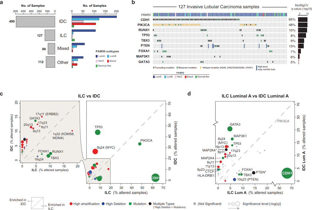
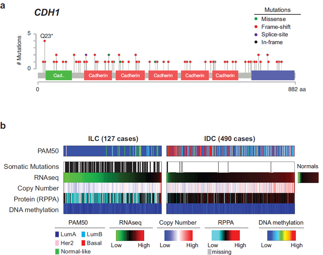
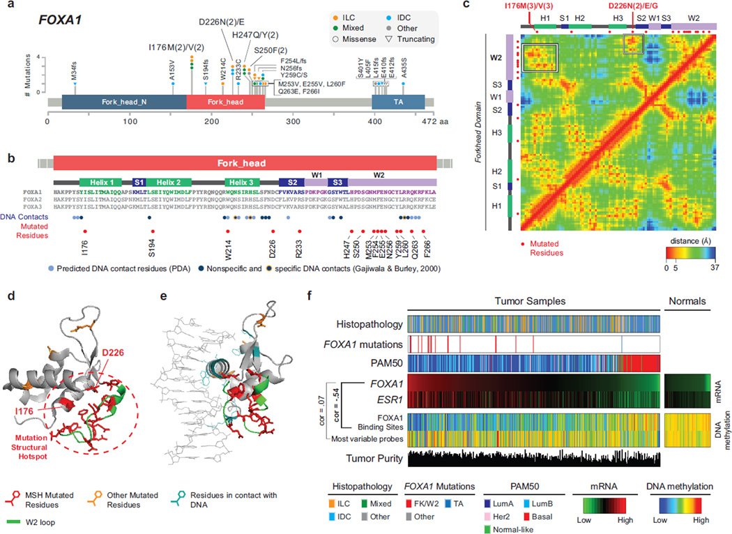
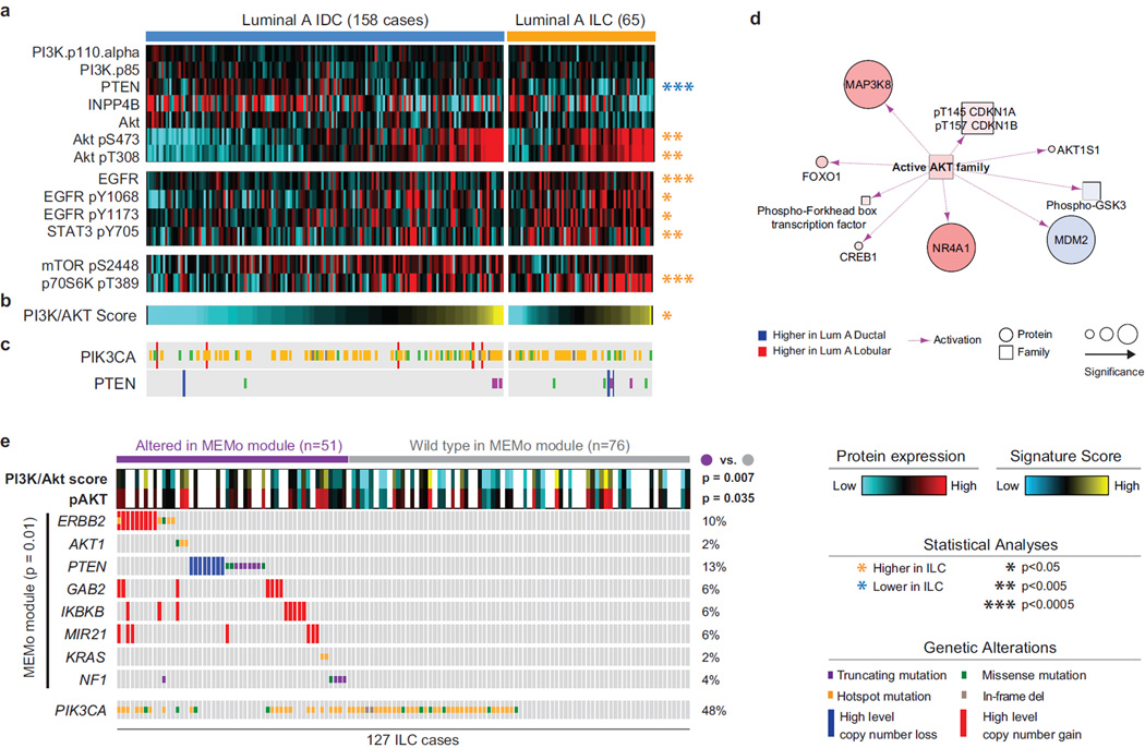
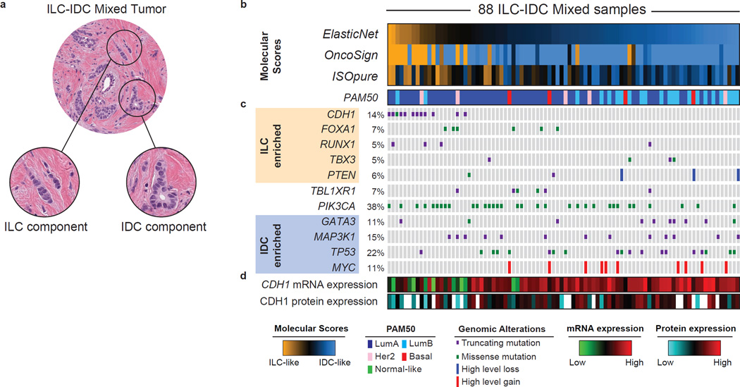
Invasive lobular carcinoma (ILC) is the second most prevalent histologic subtype of invasive breast cancer. Here, we comprehensively profiled 817 breast tumors, including 127 ILC, 490 ductal (IDC), and 88 mixed IDC/ILC. Besides E-cadherin loss, the best known ILC genetic hallmark, we identified mutations targeting PTEN, TBX3, and FOXA1 as ILC enriched features. PTEN loss associated with increased AKT phosphorylation, which was highest in ILC among all breast cancer subtypes. Spatially clustered FOXA1 mutations correlated with increased FOXA1 expression and activity. Conversely, GATA3 mutations and high expression characterized luminal A IDC, suggesting differential modulation of ER activity in ILC and IDC. Proliferation and immune-related signatures determined three ILC transcriptional subtypes associated with survival differences. Mixed IDC/ILC cases were molecularly classified as ILC-like and IDC-like revealing no true hybrid features. This multidimensional molecular atlas sheds new light on the genetic bases of ILC and provides potential clinical options.


Figures

Figure 1
Figure 1. Molecular determinants of invasive lobular breast cancer
A) Histopathological breast cancer subtypes: invasive ductal (IDC), invasive lobular (ILC), mixed ductal/lobular (Mixed), and other-type (Other) carcinoma. PAM50 intrinsic subtypes are not equally distributed across breast cancer subtypes. B) Recurrently mutated genes (MutSigCV2) in ILC. C) Comparison of the alteration frequency for 153 recurrent genomic alterations in ILC versus IDC. D) Comparison of the alteration frequency for 153 recurrent genomic alterations in ILC LumA versus IDC LumA.
Figure 2
Figure 2. E-cadherin loss in ILC
A) Mutations targeting the CDH1 gene target residues across the whole sequence and are mostly predicted to be truncating (red). B) Comparison of E-cadherin status between ILC and IDC reveals frequent hemizygous copy number losses at the CDH1 locus and downregulation of both mRNA [log2(RSEM)] and protein levels. See also Figure S2A–C. Average DNA methylation level of 6 probes at the CDH1 promoter shows no change in DNA methylation in both ILC and IDC samples. See also Figure S2D–I.
Figure 3
Figure 3. Recurrent FOXA1 mutations cluster in the 3D space and correlate with high FOXA1 activity
A) Recurrent FOXA1 mutations in 817 breast tumors cluster in the Fork-head DNA binding (FK) domain and in the C-terminus trans-activation (TA) domain. B) Secondary structure elements of the FK domain are not equally mutated. FOXA1 mutations cluster in the W2 loop and rarely target residues interacting with the DNA. C) Residue-residue minimum distances for all residues in the FK domain using the 3D structure of FOXA3 FK domain (PDB ID: 1VTN). Frequently mutated residues I176 and D226 are close in the 3D space (but not in the sequence) to the residues in the W2 loop. See also Figure S3C. D) 3D structure of the FK domain. Mutations in the W2 loop, in I176, and in D226 form a mutational structural hotspot (MSH). E) 3D structure of FK domain bound to the DNA molecule shows mutated residues (red) are not those in contact with the DNA (light blue). F) Across all breast cancer subtypes (histopathology and PAM50), FOXA1 mutations are associated with FOXA1 high mRNA expression. FOXA1 mRNA expression is highly correlated with ER mRNA expression [log−2(RSEM)] and anti-correlated with DNA methylation at FOXA1 binding sites consistent with FOXA1 activity. DNA methylation of randomly selected probes was used as control.
Figure 4
Figure 4. Akt signaling is highest in ILC tumors
A) Differential protein and phospho-protein analysis between ILC LumA and IDC LumA reveals significant lower levels of PTEN, and higher levels of Akt, phospho-Akt, EGFR, phospho-EGFR, phopsho-STAT3, and phospho-p70S6K in ILC LumA. B) A PI3K/Akt protein expression signature is significantly up-regulated in ILC tumors. See also Figure S4B–C. C) Mutation and copy number alterations in PIK3CA and PTEN D) PARADIGM identifies increased Akt activity in LumA ILC tumors. E) MEMo identified multiple mutually exclusive alterations in ILC converging on Akt signaling and associated with increased phospho-Akt and PI3K/Akt protein signature in these tumors. Hotspot are defined as follow: PIK3CA E542, E545, Q546, and H1047; ERBB2 L755, I767, V777; AKT1 E17; KRAS G12.
Figure 5
Figure 5. ILC molecular subtypes
A) Three molecular subtype of lobular breast cancer were identified based on differential gene expression and show unique patterns highly expressed genes (n=1277, SAM FDR=0, upper panel), minor difference in tumor purity measured by ABSOLUTE, and differences in gene expression signatures measuring proliferation, CD68, Macrophage-associated CSF1, Macrophage–associated TH1, and T Cell Receptor Signaling (lower panel). Proliferation is highest in the proliferative (Pro) and immune-related (IR) subgroups; macrophage associated signaling is highest in immune-related tumors. B) Differences in protein expression profiles as determined by RPPA analysis. The reactive-like (RL) subgroup shows a significant association (p<1E-4, Fisher’s Exact test) with the RPPA-defined Reactive subgroup of breast cancer. Differences in subgroup-specific patterns of protein expression (p<0.05, t-test) for individual proteins (upper panel) as well as for protein expression signatures (lower panel) were identified. The proliferative subgroup shows higher expression of the cell cycle and DNA damage response pathways and lower levels of Ras-MAPK signaling (p<0.05). C) Subgroup-associated signaling features identified by PARADIGM. D) Reactive-like (n=55) tumors have significantly better disease specific (p=0.038, HR: 0.47, log-rank) and E) overall survival (p=0.022, HR=0.50) compared to proliferative (n=44) tumors in the METABRIC cohort. See also Figure S5.
Figure 6
Figure 6. Molecular classification of mixed ductal/lobular carcinoma
A) Mixed ductal/lobular tumors present at the same time both a lobular and ductal component. B) We used three algorithmic approaches (ElasticNet, OncoSign, and ISOpure) to evaluate the resemblance of mixed tumors to either ILC (ILC-like) or IDC (IDC-like) based on molecular features. ILC-IDC scores are shown for all three approached at the top. See also Figure S6B–D. C) Genetic alterations enriched in ILC tumors are frequently found in ILC-like mixed cases (in particular CDH1 mutations), whereas those enriched in IDC are more frequent in IDC-like mixed cases. D) ILC-like mixed-cases are characterized by both low E-cadherin mRNA and protein level. See also Figure S6E.

References

    1. Akbani R, Ng PK, Werner HM, Shahmoradgoli M, Zhang F, Ju Z, Liu W, Yang JY, Yoshihara K, Li J, et al. A pan-cancer proteomic perspective on The Cancer Genome Atlas. Nature communications. 2014;5:3887. - PMC - PubMed
    1. Arpino G, Bardou VJ, Clark GM, Elledge RM. Infiltrating lobular carcinoma of the breast: tumor characteristics and clinical outcome. Breast cancer research : BCR. 2004;6:R149–R156. - PMC - PubMed
    1. Arps DP, Healy P, Zhao L, Kleer CG, Pang JC. Invasive ductal carcinoma with lobular features: a comparison study to invasive ductal and invasive lobular carcinomas of the breast. Breast cancer research and treatment. 2013;138:719–726. - PMC - PubMed
    1. Baay M, Brouwer A, Pauwels P, Peeters M, Lardon F. Tumor cells and tumor-associated macrophages: secreted proteins as potential targets for therapy. Clinical & developmental immunology. 2011;2011:565187. - PMC - PubMed
    1. Baca SC, Prandi D, Lawrence MS, Mosquera JM, Romanel A, Drier Y, Park K, Kitabayashi N, MacDonald TY, Ghandi M, et al. Punctuated evolution of prostate cancer genomes. Cell. 2013;153:666–677. - PMC - PubMed

Publication types

MeSH terms