. 2015 Oct 13;6(31):32039-60.
doi: 10.18632/oncotarget.5055.

PI3K/AKT/mTOR and sonic hedgehog pathways cooperate together to inhibit human pancreatic cancer stem cell characteristics and tumor growth

Affiliations

PI3K/AKT/mTOR and sonic hedgehog pathways cooperate together to inhibit human pancreatic cancer stem cell characteristics and tumor growth

Narinder Sharma et al. Oncotarget. .

Abstract

Cancer stem cells (CSCs) play major roles in cancer initiation, progression, and metastasis. It is evident from growing reports that PI3K/Akt/mTOR and Sonic Hedgehog (Shh) signaling pathways are aberrantly reactivated in pancreatic CSCs. Here, we examined the efficacy of combining NVP-LDE-225 (PI3K/mTOR inhibitor) and NVP-BEZ-235 (Smoothened inhibitor) on pancreatic CSCs characteristics, microRNA regulatory network, and tumor growth. NVP-LDE-225 co-operated with NVP-BEZ-235 in inhibiting pancreatic CSC's characteristics and tumor growth in mice by acting at the level of Gli. Combination of NVP-LDE-225 and NVP-BEZ-235 inhibited self-renewal capacity of CSCs by suppressing the expression of pluripotency maintaining factors Nanog, Oct-4, Sox-2 and c-Myc, and transcription of Gli. NVP-LDE-225 co-operated with NVP-BEZ-235 to inhibit Lin28/Let7a/Kras axis in pancreatic CSCs. Furthermore, a superior interaction of these drugs was observed on spheroid formation by pancreatic CSCs isolated from Pankras/p53 mice. The combination of these drugs also showed superior effects on the expression of proteins involved in cell proliferation, survival and apoptosis. In addition, NVP-LDE-225 co-operated with NVP-BEZ-235 in inhibiting EMT through modulation of cadherin, vimentin and transcription factors Snail, Slug and Zeb1. In conclusion, these data suggest that the combined inhibition of PI3K/Akt/mTOR and Shh pathways may be beneficial for the treatment of pancreatic cancer.

Keywords: Gli; PI3K/AKT/mTOR; cancer stem cell; pancreatic cancer; sonic hedgehog.


Conflict of interest statement

CONFLICTS OF INTEREST

All the authors of this article have declared “no conflict of interest”.

Figures

Figure 1
Figure 1. Regulation of PI3/Akt/mTOR and Shh pathways by NVP-BEZ-235, and NVP-LDE-225 in pancreatic CSCs, respectively
A. CD44+CD24+ESA+ CSCs were isolated from human primary pancreatic tumors, and treated with NVP-BEZ-235 (0–5 μM) for 7 days. At the end of incubation period, spheroids were collected, reseeded and treated with NVP-BEZ-235 for another week to obtain secondary spheroids. *, @, &, #, and ** = significantly different from control, P < 0.05. B. Pancreatic CSCs were seeded in soft agar and treated with NVP-BEZ-235 (0–5 μM) for 21 days. At the end of incubation period, numbers of colonies were counted. *, &, %, and # = significantly different from control, P < 0.05. C. Pancreatic CSCs were treated with different concentrations of NVP-BEZ-235 (0–2 μM) and expression of phospho-Akt, Phospho-PI3K, Phospho-p70S6K, Phospho-GSK3Kβ, Phospho-4EBP1, total Akt, p70S6K, total GSK3Kβ, and total 4EBP1 was analyzed by Western blot analysis. β-Actin was used as a loading control. D. Pancreatic CSCs were treated with NVP-LDE-225 (0–15 μM) for 7 days. At the end of incubation period, spheroids were collected, reseeded and treated with NVP-LDE-225 for another week to obtain secondary spheroids. *, @, &, #, and ** = significantly different from control, P < 0.05. E. Pancreatic CSCs were seeded in soft agar and treated with NVP-LDE-225 (0–15 μM) for 21 days. At the end of incubation period, numbers of colonies were counted. *, &, %, and # = significantly different from control, P < 0.05. F. CSCs were transduced with Gli-responsive GFP/firefly luciferase viral particles (pGreen Fire1-Gli with EF1, System Biosciences), and treated with NVP-LDE-225 (0–10 μM) for 36 h. Gli reporter activity was measured by luciferase assay. G. Pancreatic CSCs were treated with NVP-LDE-225 (0–10 μM) for 48 h. The expression of Gli1 and Gli2 was measured by qRT-PCR. Data represent mean ± SD. * &, and % = significantly different from control, P < 0.05. H. Effect of NVP-LDE-225 on components of Shh pathway. Pancreatic CSCs were treated with different concentrations of NVP-LDE-225 (0–10 μM) for 48 h, and expressions of Gli1, Gli2, Patched1, Patched2, smoothened and SuFu were determined by Western blot analysis. β-Actin was used as a loading control.
Figure 2
Figure 2. Effect of NVP-LDE-225, NVP-BEZ-235 and their combination on cell viability, apoptosis, and colony and spheroid formation, and protein expression
A. Effects LDE-225, BEZ-235 and their combination on cell viability. Pancreatic CSCs were seeded in 96 well plates and treated with NVP-LDE-225 (5 μM), NVP-BEZ -235 (1 μM) and their combination for 48 h. Thereafter, cell viability was measured by XTT assay. Viable cells were quantified. B. Pancreatic CSCs were treated with NVP-LDE-225 (5 μM), NVP-BEZ -235 (1 μM) and their combination for 48 h. Thereafter, cell apoptosis was measured by PI staining. Data are representative of 3 independent experiments. C. Quantification of apoptotic cells. Data represent mean (n = 4) ± S.D. *, #, and $ = significantly different from control, P < 0.05. D. Expression of cleaved caspase-3 and cleaved PARP in pancreatic CSCs treated with NVP-LDE-225 (5 μM), NVP-BEZ -235 (1 μM) and their combination. Cells were treated with these drug(s) for 48 h, and the expression of cleaved caspase-3 and PARP was analyzed by Western blot analysis. E. Pancreatic CSCS were treated with NVP-LDE-225, NVP-BEZ-235 and their combination for 48 h, thereafter, fresh media added to cells. After 10 days, colonies were observed by staining the plates with 0.5% crystal violet strain. F. Quantification of pancreatic CSCs colonies. Data represent mean (n = 4) ± S.D. *, #, and $ = significantly different from control, P < 0.05. G. Effects of NVP-LDE-225, and/or NVP-BEZ -235 on spheroid formation by pancreatic CSC. Pancreatic CSCs were seeded in suspension and treated with NVP-LDE-225 (5 μM), NVP-BEZ -235 (1 μM) and their combination for 7 days. Spheroid images were obtained by light microscopy. H. qRT-PCR analysis of expression profile for Bcl-2, XIAP, BMI and CCND1 in pancreatic CSCs after treatment with NVP-LDE-225 (5 μM), NVP-BEZ -235 (1 μM) and their combination for 36 h. HK-GAPD was used as the endogenous normalization control. Data represent mean (n = 4) ± S.D. *, #, and @ = significantly different from control, P < 0.05. I. Pancreatic CSCs were treated with NVP-LDE-225, NVP-BEZ-235 and their combination for 48 h, and the expression of Bcl2, Bcl-XL, cIAP1, cIAP2, Bim, cyclin D1, and β-actin (loading control) was measured by Western blot analysis. J. Pancreatic CSCs were transduced with Gli-responsive GFP/firefly luciferase viral particles (pGreen Fire1-Gli with EF1, System Biosciences). After transduction, culture medium was replaced and CSCs were treated with BEZ235 (1 μM) and/or LDE225 (5 μM) for 24 h. Gli reporter activity was measured as we described [82]. K. Pancreatic CSCs were treated with NVP-LDE-225 (5 μM), NVP-BEZ-235 (1 μM) or their combination for 36 h. RNA was extracted and expressions of Gli1, Gli2, Patched1 (PTCH1), and Patched2 (PTCH2) were measured by qRT-PCR. Data represent mean (n = 4) ± S.D. *, #, and @ = significantly different from control, P < 0.05.
Figure 3
Figure 3. NVP-LDE-225, NVP-BEZ-235 and their combination inhibit spheroid formation by CSCs isolated from pancreas of human and KrasG12D; Trp53LSL-R172H/+ PDAC (Pankras/p53) mice, and differentially regulates genes involved in self-renewal and pluripotency of pancreatic CSCs
A. Human pancreatic CSCs isolated from primary tumors were treated with NVP-LDE-225, NVP-BEZ-235 and their combination for 7 days to obtain primary spheroids. At the end of incubation period, spheroids were collected, reseeded and treated with NVP-LDE-225 and/or NVP-BEZ-235 for another week to obtain secondary spheroids. Secondary spheroids were collected, reseeded and treated with NVP-LDE-225 and/or NVP-BEZ-235 for another week to obtain tertiary spheroids. Cell viability in spheroids was measured by trypan blue assay at the end of 7, 14 and 21 days. Data represent mean ± SD. *, &, #, % and $ = significantly different from control, P < 0.05. B. Pancreatic CSCs isolated from Pankras/p53 mice were treated with NVP-LDE-225, NVP-BEZ-235 and their combination for 7 days to obtain primary spheroids. At the end of incubation period, spheroids were collected, reseeded and treated with NVP-LDE-225 and/or NVP-BEZ-235 for another week to obtain secondary spheroids. Secondary spheroids were collected, reseeded and treated with NVP-LDE-225 and/or NVP-BEZ-235 for another week to obtain tertiary spheroids. Cell viability in spheroids was measured by trypan blue assay at the end of 7, 14 and 21 days. Data represent mean ± SD. *, &, #, % and $ = significantly different from control, P < 0.05. C. Pancreatic CSCs were treated with NVP-LDE-225, NVP-BEZ-235 and their combination for 36 h, and expressions of Nanog, Oct-4, c-Myc and Sox-2 were quantified by qRT-PCR. HK-GAPDH was used as the endogenous normalization control. Data represent mean (n = 4) ± SD. * = significant difference from control, P < 0.05. $ = significant difference from control or single agent alone, P < 0.05. D. Protein expression of Nanog, c-Myc, Oct-4 and Sox-2. Pancreatic CSCs were treated with NVP-LDE-225, NVP-BEZ-235 and their combination for 48 h, and the expression of Nanog, c-Myc, Oct-4 and Sox-2 was determined by the Western blot analysis. β-actin was used as a loading control. E. and F. Immunohistochemical examination of Nanog, Oct-4, c-Myc, and Sox2 in pancreatic CSCs treated with NVP-LDE-225, NVP-BEZ-235 and their combination. Pancreatic CSCs were grown in suspension and treated with above mentioned drugs for 48 h. Spheroids formed by pancreatic CSCs were fixed and IHC was performed as described in Material and Methods.
Figure 4
Figure 4. NVP-LDE-225, NVP-BEZ-235 and their combination regulate the pancreatic CSC motility and migration, and the expression of EMT-related genes
A. Motility Assay. In vitro motility of pancreatic CSCs was observed by using scratch technique. CSCs were grown in monolayer, scratched and treated with NVP-LDE-225, NVP-BEZ-235 and their combination for 48 h. Data are representative of three independent experiments. B. Transwell migration assay. Pancreatic CSCs were plated in the top chamber of the transwell and treated with NVP-LDE-225, NVP-BEZ-235 and their combination for 24 h. cells migrated to the lower chambered were fixed with methanol, stained with crystal violet and counted. Data represent mean (n = 4) ± S.D. * = significantly different from control, P < 0.05. $ = significantly different from control or single drug treatment, P < 0.05. C–E. Pancreatic CSCs were treated with NVP-LDE-225, NVP-BEZ-235 and their combination for 36 h. After incubation, the expression of snail, slug and Zeb1 was measured by qRT–PCR. Data represent mean (n = 4) ± S.D. * = significantly different from control, P < 0.05. $ = significantly different from control or single drug treatment, P < 0.05.
Figure 5
Figure 5. Regulation of miRNAs in human pancreatic CSCs treated with NVP-BEZ-235 and/or NVP-LDE-225
A. Pancreatic CSCs were treated with NVP-BEZ-235 (1 μM) for 36 h. RNA was isolated and miRNA array (Affimetrix gene Chip) analysis was performed. Expression of selected miRNAs. P-values and fold change are also shown. B. Bio-function analysis. C. Netwrok analysis. D. Pancreatic CSCs were treated with NVP-LDE-225 (5 μM) for 36 h. RNA was isolated and miRNA array (Affimetrix gene Chip) analysis was performed. Expression of selected miRNAs. P-values and fold change are also shown. E. Bio-function analysis. F. Netwrok analysis. G. Pancreatic CSCs were treated with NVP-BEZ-235 (1 μM) and NVP-LDE-225 (5 μM) for 36 h. RNA was isolated and miRNA array (Affimetrix gene Chip) analysis was performed. Left panel, Expression of selected miRNAs. P-values and fold change are also shown. Right panel, Heat Map of miRNA arrays from NVP-LDE-225- and/or NVP-BEZ-235- treated pancreatic CSCs. H. Bio-function analysis. I. Netwrok analysis.
Figure 6
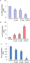
Figure 6. BEZ235 and LDE225 regulate Lin28/Let7a/Kras axis in pancreatic CSCs
A–C. Expression of Lin28a, Let7a and Kras. Pancreatic CSCs were treated with BEZ235 (1 μM) and/or LDE225 (5 μM) for 36 h. RNA was extracted and the expression of Lin28a, Let7a and Kras was measured by qRT-PCR. Data represent mean (n = 4) ± S.D. * and $ = significantly different from control, P < 0.05.
Figure 7
Figure 7. NVP-LDE-225, NVP-BEZ-235 and their combination inhibit pancreatic CSC tumor growth in NOD/SCID IL2Rγ null mice
A. Effects of NVP-LDE-225 and/or NVP-BEZ-235 on pancreatic CSC tumor growth in NOD/SCID IL2R γ null mice. Tumor-bearing nude mice were injected with vehicle or NVP-LDE-225 (40 mg/kg), NVP-BEZ-235 (20 mg/kg) and their combination intraperitoneally, five times per week for 4 weeks. Tumor weights were measured at the end of the experiments. Data represent mean ± S.D. * = significantly different from control, P < 0.05. $ = significantly different from control or single drug treatment, P < 0.05. B. Expression of pluripotency maintaining factors. Tissue lysates were prepared from tumor tissues isolated from control and NVP-LDE-225, NVP-BEZ-235 and their combination treated mice. Western blot analysis was performed to measure the expression of c-Myc, Sox-2, Nanog, and Oct-4. C. Expression of Bcl-2 family members. Tissue lysates were prepared and the expression of Bcl2, Bad, Bax, and Bak was measured by the Western blot analysis. β-actin was used as a loading control. D. Expression of Caspase 3 and Caspase 9. Tissue lysates were prepared and the expression of Caspase 3 and Caspase 9 was measured by the Western blot analysis. β-actin was used as a loading control. E. Expression of E-cadherin, Vimentin, Slug, and Snail. Tissue lysates were prepared and the expression of E-cadherin, Vimentin, Slug, and Snail was measured by the Western blot analysis. β-actin was used as a loading control.

Similar articles

Cited by

References

    1. Iovanna J, Neira JL. Pancreatic cancer: molecular, biochemical, chemopreventive, and therapeutic aspects. The Scientific World Journal. 2010;10:1967–1970. - PMC - PubMed
    1. Siegel R, Ma J, Zou Z, Jemal A. Cancer statistics, 2014. CA: a cancer journal for clinicians. 2014;64:9–29. - PubMed
    1. Adhikari AS, Agarwal N, Iwakuma T. Metastatic potential of tumor-initiating cells in solid tumors. Front Biosci. 2011;16:1927–1938. - PubMed
    1. Wang Z, Li Y, Ahmad A, Banerjee S, Azmi AS, Kong D, Sarkar FH. Pancreatic cancer: understanding and overcoming chemoresistance. Nature reviews Gastroenterology & hepatology. 2011;8:27–33. - PubMed
    1. Nusslein-Volhard C, Wieschaus E. Mutations affecting segment number and polarity in Drosophila. Nature. 1980;287:795–801. - PubMed

MeSH terms