

Role of N-cadherin in proliferation, migration, and invasion of germ cell tumours
- PMID: 26451610
- PMCID: PMC4741776
- DOI: 10.18632/oncotarget.5288
Role of N-cadherin in proliferation, migration, and invasion of germ cell tumours
Abstract
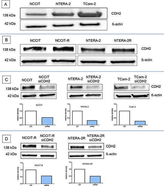
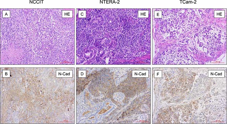
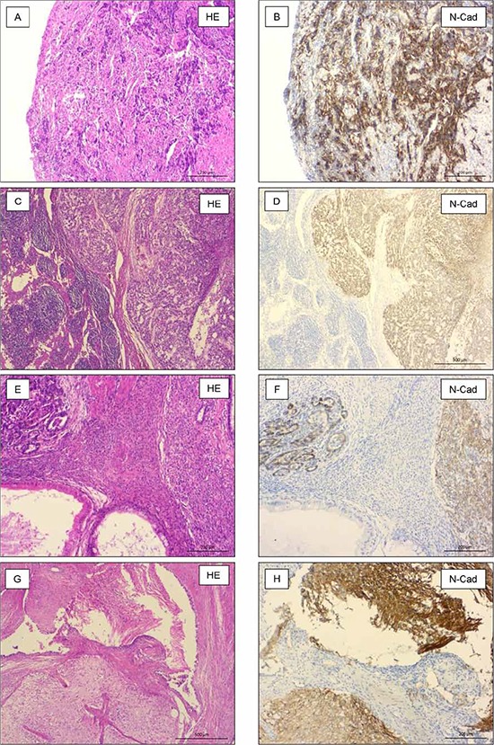
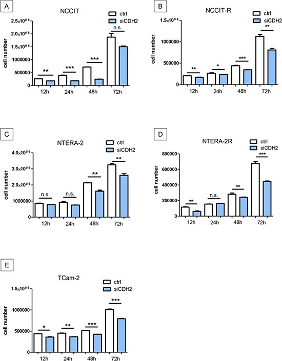
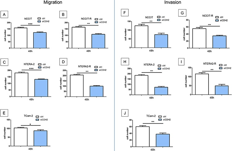
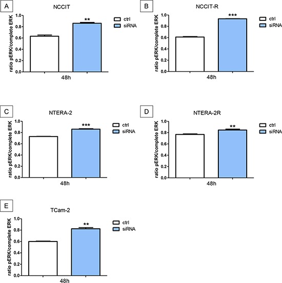
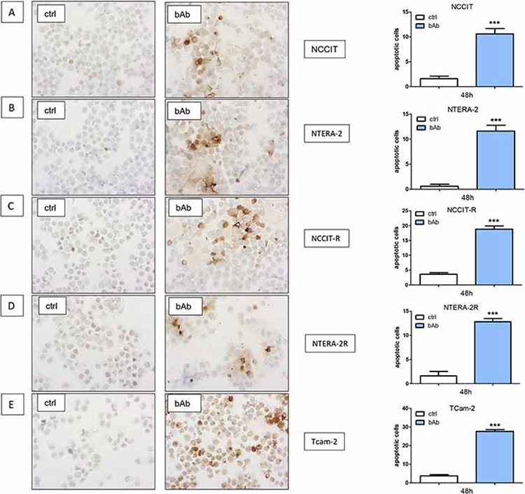
Germ cell tumors (GCTs) are the most common malignancies in young men. Most patients with GCT can be cured with cisplatin-based combination chemotherapy, even in metastatic disease. In case of therapy resistance, prognosis is usually poor. We investigated the potential of N-cadherin inhibition as a therapeutic strategy. We analyzed the GCT cell lines NCCIT, NTERA-2, TCam-2, and the cisplatin-resistant sublines NCCIT-R and NTERA-2R. Effects of a blocking antibody or siRNA against N-cadherin on proliferation, migration, and invasion were investigated. Mouse xenografts of GCT cell lines were analyzed by immunohistochemistry for N-cadherin expression. All investigated GCT cell lines were found to express N-cadherin protein in vitro and in vivo. Downregulation of N-cadherin in vitro leads to a significant inhibition of proliferation, migration, and invasion. N-cadherin-downregulation leads to a significantly higher level of pERK. N-cadherin-inhibition resulted in significantly higher rates of apoptotic cells in caspase-3 staining. Expression of N-cadherin is preserved in cisplatin-resistant GCT cells, pointing to an important physiological role in cell survival. N-cadherin-downregulation results in a significant decrease of proliferation, migration, and invasion and stimulates apoptosis in cisplatin-naive and resistant GCT cell lines. Therefore, targeting N-cadherin may be a promising therapeutic approach, particularly in cisplatin-resistant, therapy refractory and metastatic GCT.
Keywords: GCT-cell lines; N-cadherin; cisplatin resistance; germ cell tumours.
Conflict of interest statement
The authors declare that they have no competing interests.
Figures
Similar articles
-
Mirvetuximab Soravtansine Induces Potent Cytotoxicity and Bystander Effect in Cisplatin-Resistant Germ Cell Tumor Cells.Cells. 2025 Feb 15;14(4):287. doi: 10.3390/cells14040287. Cells. 2025. PMID: 39996761 Free PMC article.
-
Targeting of Deregulated Wnt/β-Catenin Signaling by PRI-724 and LGK974 Inhibitors in Germ Cell Tumor Cell Lines.Int J Mol Sci. 2021 Apr 20;22(8):4263. doi: 10.3390/ijms22084263. Int J Mol Sci. 2021. PMID: 33923996 Free PMC article.
-
Increased levels of XPA might be the basis of cisplatin resistance in germ cell tumours.BMC Cancer. 2020 Jan 6;20(1):17. doi: 10.1186/s12885-019-6496-1. BMC Cancer. 2020. PMID: 31906898 Free PMC article.
-
Third-line chemotherapy and novel agents for metastatic germ cell tumors.Hematol Oncol Clin North Am. 2011 Jun;25(3):577-91, ix. doi: 10.1016/j.hoc.2011.03.005. Epub 2011 Apr 22. Hematol Oncol Clin North Am. 2011. PMID: 21570610 Review.
-
Cisplatin resistance in germ cell tumours: models and mechanisms.Andrology. 2015 Jan;3(1):111-21. doi: 10.1111/andr.299. Epub 2014 Dec 29. Andrology. 2015. PMID: 25546083 Review.
Cited by
-
Combined targeting of TCF7L1/2, PTEN, CDK6, and BCCIP by microRNA miR-29c-3p is associated with reduced invasion and proliferation of endometriotic cells.Reprod Med Biol. 2025 Mar 25;24(1):e12645. doi: 10.1002/rmb2.12645. eCollection 2025 Jan-Dec. Reprod Med Biol. 2025. PMID: 40135061 Free PMC article.
-
Proteomic Comparison of Malignant Human Germ Cell Tumor Cell Lines.Dis Markers. 2019 Sep 3;2019:8298524. doi: 10.1155/2019/8298524. eCollection 2019. Dis Markers. 2019. PMID: 31565104 Free PMC article.
-
Alterations in androgen deprivation enhanced prostate-specific membrane antigen (PSMA) expression in prostate cancer cells as a target for diagnostics and therapy.EJNMMI Res. 2015 Dec;5(1):66. doi: 10.1186/s13550-015-0145-8. Epub 2015 Nov 17. EJNMMI Res. 2015. PMID: 26576996 Free PMC article.
-
Cisplatin‑resistant germ cell tumor models: An exploration of the epithelial‑mesenchymal transition regulator SLUG.Mol Med Rep. 2024 Dec;30(6):228. doi: 10.3892/mmr.2024.13352. Epub 2024 Oct 11. Mol Med Rep. 2024. PMID: 39392037 Free PMC article.
-
Human giant larvae-1 promotes migration and invasion of malignant glioma cells by regulating N-cadherin.Oncol Lett. 2021 Feb;21(2):167. doi: 10.3892/ol.2021.12428. Epub 2021 Jan 4. Oncol Lett. 2021. PMID: 33552285 Free PMC article.
References
-
- Beyer J, Albers P, Altena R, Aparicio J, Bokemeyer C, Busch J, Cathomas R, Cavallin-Stahl E, Clarke NW, Classen J, Cohn-Cedermark G, Dahl AA, Daugaard G, De Giorgi U, De Santis M, De Wit M, et al. Maintaining success, reducing treatment burden, focusing on survivorship: highlights from the third European consensus conference on diagnosis and treatment of germ-cell cancer. Annals of oncology : official journal of the European Society for Medical Oncology / ESMO. 2013;24:878–888. - PMC - PubMed
-
- Horwich A, Shipley J, Huddart R. Testicular germ-cell cancer. Lancet. 2006;367:754–765. - PubMed
-
- Hoei-Hansen CE, Nielsen JE, Almstrup K, Sonne SB, Graem N, Skakkebaek NE, Leffers H, Rajpert-De Meyts E. Transcription factor AP-2gamma is a developmentally regulated marker of testicular carcinoma in situ and germ cell tumors. Clinical cancer research : an official journal of the American Association for Cancer Research. 2004;10:8521–8530. - PubMed
-
- Bokemeyer C, Oechsle K, Honecker F, Mayer F, Hartmann JT, Waller CF, Bohlke I, Kollmannsberger C. Combination chemotherapy with gemcitabine, oxaliplatin, and paclitaxel in patients with cisplatin-refractory or multiply relapsed germ-cell tumors: a study of the German Testicular Cancer Study Group. Annals of oncology : official journal of the European Society for Medical Oncology / ESMO. 2008;19:448–453. - PubMed
-
- Feldman DR, Bosl GJ, Sheinfeld J, Motzer RJ. Medical treatment of advanced testicular cancer. Jama. 2008;299:672–684. - PubMed
Publication types
MeSH terms
Substances
LinkOut - more resources
Full Text Sources
Other Literature Sources
Medical
Research Materials
