

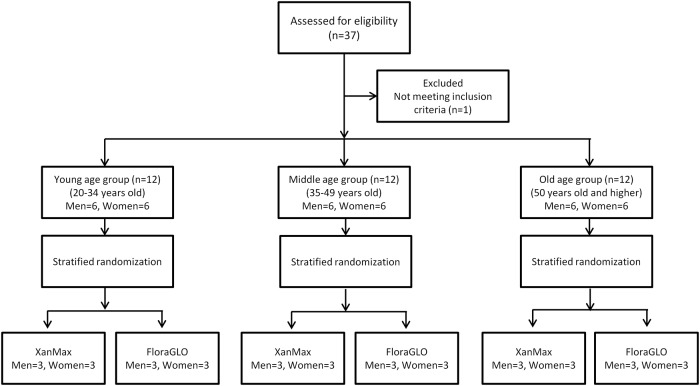
Changes in Macular Pigment Optical Density and Serum Lutein Concentration in Japanese Subjects Taking Two Different Lutein Supplements
- PMID: 26451726
- PMCID: PMC4599964
- DOI: 10.1371/journal.pone.0139257
Changes in Macular Pigment Optical Density and Serum Lutein Concentration in Japanese Subjects Taking Two Different Lutein Supplements
Abstract
Purpose: To investigate macular pigment optical density (MPOD) and serum concentration changes of lutein in Japanese subjects participating in a clinical trial in which two formulations of lutein and zeaxanthin supplements with different physiochemical properties are used.
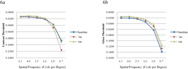
Methods: Thirty-six healthy volunteers were recruited into this prospective, randomized, parallel-group, double-masked comparative study at a single institute. Two products were used, FloraGLO® (Kemin Japan) and XanMax® (Katra Phytochem). The lutein particle size and zeaxanthin concentrations differed between the formulations. The subjects consumed one of the two supplements for a duration of up to 6 months. MPOD levels were measured by resonance Raman spectrometry at baseline and once a month until the end of the study. Serum lutein concentration was measured at baseline, month 3, and month 6. The subjects were also tested for contrast sensitivity, glare sensitivity, visual acuity, and in addition had a focal electroretinogram measured.
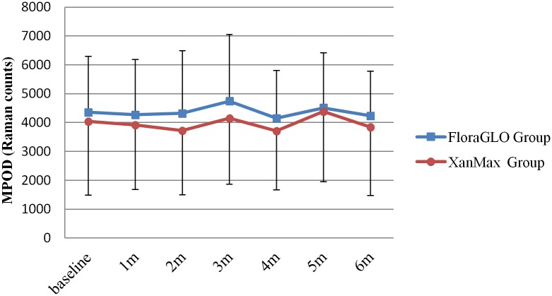
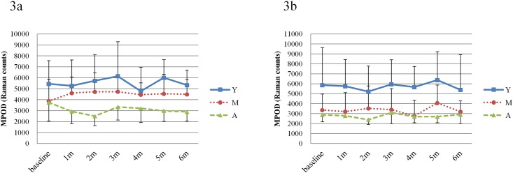
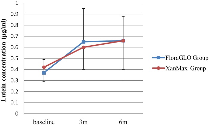
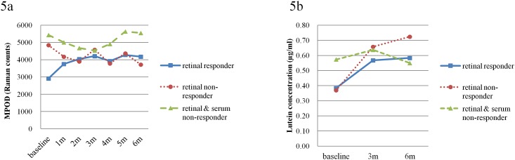
Results: The mean serum lutein concentrations increased significantly after the first three months, but the mean MPOD levels in either supplement group did not show any statistically significant increase. A detailed analysis, however, revealed three response patterns in both groups for the increase of MPOD levels and serum lutein concentration, i.e. "retinal responders", who had an increase of both MPOD levels and serum lutein concentrations (n = 13), "retinal non-responders", who had only increased serum concentrations and no change in MPOD levels (n = 20), and "retinal and serum non-responders", who had neither MPOD level nor plasma concentration increases (n = 3). The subjects with low MPOD levels at baseline appeared to show increased MPOD levels at the 6 month time point upon lutein supplementation (r = -0.4090, p = 0.0133). Glare sensitivity improved in retinal responders in both supplement groups, while there were no remarkable changes in contrast sensitivity.
Conclusions: No statistically significant differences could be detected for MPOD levels and serum lutein concentrations between the two investigated lutein supplement formulations. Responses to lutein supplementation regarding MPOD levels and serum lutein concentrations varied between subjects. Subjects with lower MPOD levels at baseline responded well to lutein supplementation. However, since the number of subjects was low, a further study with more subjects is needed to prove that subjects with low MPOD levels will benefit from lutein supplementation.
Trial registration: UMIN-CTR UMIN000004593.
Conflict of interest statement
Figures
Similar articles
-
The Effect of Lutein/Zeaxanthin Intake on Human Macular Pigment Optical Density: A Systematic Review and Meta-Analysis.Adv Nutr. 2021 Dec 1;12(6):2244-2254. doi: 10.1093/advances/nmab071. Adv Nutr. 2021. PMID: 34157098 Free PMC article.
-
Serum and retinal responses to three different doses of macular carotenoids over 12 weeks of supplementation.Exp Eye Res. 2016 Oct;151:1-8. doi: 10.1016/j.exer.2016.07.005. Epub 2016 Jul 15. Exp Eye Res. 2016. PMID: 27426932 Clinical Trial.
-
Macular pigment density variation after supplementation of lutein and zeaxanthin using the Visucam® 200 pigment module: Impact of age-related macular degeneration and lens status.J Fr Ophtalmol. 2017 Apr;40(4):303-313. doi: 10.1016/j.jfo.2016.11.009. Epub 2017 Mar 21. J Fr Ophtalmol. 2017. PMID: 28336284 Clinical Trial.
-
Comparison of macular pigment and serum lutein concentration changes between free lutein and lutein esters supplements in Japanese subjects.Acta Ophthalmol. 2016 Sep;94(6):e411-6. doi: 10.1111/aos.13106. Epub 2016 Jun 8. Acta Ophthalmol. 2016. PMID: 27273910 Clinical Trial.
-
Lutein, Zeaxanthin and Meso-zeaxanthin Supplementation Associated with Macular Pigment Optical Density.Nutrients. 2016 Jul 12;8(7):426. doi: 10.3390/nu8070426. Nutrients. 2016. PMID: 27420092 Free PMC article. Review.
Cited by
-
Macular Pigment Optical Density and Photoreceptor Outer Segment Length as Predisease Biomarkers for Age-Related Macular Degeneration.J Clin Med. 2020 May 5;9(5):1347. doi: 10.3390/jcm9051347. J Clin Med. 2020. PMID: 32380638 Free PMC article.
-
Effect of Antioxidant Supplementation on Macular Pigment Optical Density and Visual Functions: A Systematic Review and Network Meta-Analysis of Randomized Controlled Trials.Adv Nutr. 2024 May;15(5):100216. doi: 10.1016/j.advnut.2024.100216. Epub 2024 Apr 4. Adv Nutr. 2024. PMID: 38582248 Free PMC article.
-
The Effect of Lutein/Zeaxanthin Intake on Human Macular Pigment Optical Density: A Systematic Review and Meta-Analysis.Adv Nutr. 2021 Dec 1;12(6):2244-2254. doi: 10.1093/advances/nmab071. Adv Nutr. 2021. PMID: 34157098 Free PMC article.
-
The Effect of Lutein on Eye and Extra-Eye Health.Nutrients. 2018 Sep 18;10(9):1321. doi: 10.3390/nu10091321. Nutrients. 2018. PMID: 30231532 Free PMC article. Review.
-
Effects of Macuprev® Supplementation in Age-Related Macular Degeneration: A Double-Blind Randomized Morpho-Functional Study Along 6 Months of Follow-Up.Adv Ther. 2019 Sep;36(9):2493-2505. doi: 10.1007/s12325-019-01016-2. Epub 2019 Jun 25. Adv Ther. 2019. PMID: 31243641 Free PMC article. Clinical Trial.
References
-
- Bone RA, Landrum JT, Hime GW, Cains A, Zamor J. Stereochemistry of the human macular carotenoids. Invest Ophthalmol Vis Sci. 1993;34:2033–2040. - PubMed
-
- Landrum JT, Bone RA. Lutein, zeaxanthin, and the macular pigment. Arch Biochem Biophys. 2001; 385: 28–40. - PubMed
-
- Krinsky NI, Landrum JT, Bone RA. Biologic mechanisms of the protective role of lutein and zeaxanthin in the eye. Annu Rev Nutr. 2003; 23: 171–201. - PubMed
-
- Krinsky NI, Johnson EJ. Carotenoid actions and their relation to health and disease. Mol Aspects Med. 2005; 26: 459–516. - PubMed
-
- Ritcher SP, Stiles W, Statkute L, Pulido J, Frankowski J, Rudy D, et al. Double-masked, placebo-controlled, randomized trial of lutein and antioxidant supplementation in the intervention of atrophic age-related macular degeneration: the Veterans LAST study (Lutein Antioxidant Supplementation Trial). Optometry 2004;75:216–230. - PubMed
Publication types
MeSH terms
Substances
Associated data
Grants and funding
LinkOut - more resources
Full Text Sources
Other Literature Sources
Medical
