. 2016 Jan;147(1):55-72.
doi: 10.1111/imm.12541. Epub 2015 Nov 6.

Contribution of Fcγ receptors to human respiratory syncytial virus pathogenesis and the impairment of T-cell activation by dendritic cells

Affiliations

Contribution of Fcγ receptors to human respiratory syncytial virus pathogenesis and the impairment of T-cell activation by dendritic cells

Roberto S Gómez et al. Immunology. 2016 Jan.

Abstract

Human respiratory syncytial virus (hRSV) is the leading cause of infant hospitalization related to respiratory disease. Infection with hRSV produces abundant infiltration of immune cells into the airways, which combined with an exacerbated pro-inflammatory immune response can lead to significant damage to the lungs. Human RSV re-infection is extremely frequent, suggesting that this virus may have evolved molecular mechanisms that interfere with host adaptive immunity. Infection with hRSV can be reduced by administering a humanized neutralizing antibody against the virus fusion protein in high-risk infants. Although neutralizing antibodies against hRSV effectively block the infection of airway epithelial cells, here we show that both, bone marrow-derived dendritic cells (DCs) and lung DCs undergo infection with IgG-coated virus (hRSV-IC), albeit abortive. Yet, this is enough to negatively modulate DC function. We observed that such a process is mediated by Fcγ receptors (FcγRs) expressed on the surface of DCs. Remarkably, we also observed that in the absence of hRSV-specific antibodies FcγRIII knockout mice displayed significantly less cellular infiltration in the lungs after hRSV infection, compared with wild-type mice, suggesting a potentially harmful, IgG-independent role for this receptor in hRSV disease. Our findings support the notion that FcγRs can contribute significantly to the modulation of DC function by hRSV and hRSV-IC. Further, we provide evidence for an involvement of FcγRIII in the development of hRSV pathogenesis.

Keywords: Fcγ receptors; dendritic cells; human respiratory syncytial virus; immune complexes; neutralizing antibodies; palivizumab.


Figures

Figure 1
Figure 1
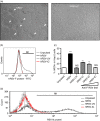
Human respiratory syncytial virus (hRSV) coated with an IgG1 neutralizing antibody displays impaired capacity to infect HEp‐2 cells. HEp‐2 cells were inoculated with hRSV and IgG‐coated hRSV (hRSVIC) at a multiplicity of infection (MOI) of 1 for 48 hr and then analysed for RSV infection. (a) Cytopathic effect (syncytia formation, arrowheads) on HEp‐2 cells inoculated with hRSV and hRSVIC. Images were taken at 40 × magnification. (b) Expression of hRSV F protein on the surface of HEp‐2 cells determined by flow cytometry. Representative histogram showing the expression of F protein on the surface of HEp‐2 cells pulsed either with hRSV (black), hRSVUV (grey) or hRSVIC (red). Uninfected cells were included as a control (thin black line). (c) Quantification of HEp‐2 cells positive for F protein expression after infection with hRSV, hRSVUV and hRSVIC. The hRSVIC was prepared with increasing amounts of a monoclonal neutralizing antibody. (d) FACS analysis of intracellular N protein expression in HEp‐2 cells pulsed with hRSV (black), hRSVUV (grey) and hRSVIC (red) using an antibody dilution equal to 0·86 mg/ml with hRSV. Data are means ± SEM of two independent experiments **P < 0·01, ***P < 0·001; data were analysed by one‐way analysis of variance and Bonferroni post‐test.
Figure 2
Figure 2
IgG‐coated human respiratory syncytial virus (hRSVIC) infects murine dendritic cells (DCs). DCs derived from wild‐type (WT), Fcγ RIIb−/− and Fcγ RIII −/− mice were incubated overnight with hRSV at a multiplicity of infection (MOI) equal to 1 and analysed 48 hr later for hRSV infection. Pre‐treatment with the Fc‐blocking antibody 2.4G2 was applied when indicated. (a) Gate strategy used for analysing hRSV‐infected DCs. CD11c+ cells were subsequently analysed on histogram overlays for F protein expression. The M1 marker was used to measure F‐derived fluorescence beyond the baseline of unstained cells. The grey‐filled histogram represents control CD11c+ cells stained with the secondary antibody. (b) Expression of RSV F protein on the surface of CD11c+ DCs pulsed either with hRSV or hRSVIC measured by FACS. Uninfected DCs were included as control. (c) Real‐time quantitative PCR for the detection of RSV nucleoprotein RNA in DCs. Graphs show number of copies of N RNA molecules per 5000 copies of β‐actin. (d) Generation of infective hRSV particles by plaque assay on HEp‐2 cells. As a control we included supernatants of uninfected or hRSV‐infected HEp‐2 cells. Plaque‐forming units per ml (PFU/ml) from supernatants of hRSV and hRSVIC inoculated. Data are means ± SEM of three to nine independent experiments *P < 0·05, **P < 0·01, ***P < 0·001, ns: non‐significant; data were analysed by one‐way analysis of variance and Bonferroni post‐test.
Figure 3
Figure 3
IgG‐coated human respiratory syncytial virus (hRSVIC) impairs the capacity of dendritic cells (DCs) to activate naive T cells. (a, c and e) Secretion of interleukin‐2 (IL‐2) by OTII CD4+ and (b, d and f) interferon‐γ (IFNγ) by OT‐I CD8+ T cells stimulated with pOVA‐pulsed DCs (a and b wild‐type (WT) DCs: c and d Fcγ RIIb−/− DCs; and e and f Fcγ RIII −/− DCs), either uninfected, hRSV‐ or hRSVIC‐inoculated. When indicated, DCs were pre‐treated with 2.4G2 to block Fcγ RIIB and Fcγ RIII. Data are means ± SEM of at least three independent experiments *P < 0·05, **P < 0·01, ***P < 0·001, ns: non‐significant; data were analysed by one‐way analysis of variance and Bonferroni post‐test.
Figure 4
Figure 4
Pulmonary dendritic cell (DC) infection and in vivo T‐cell responses after human respiratory syncytial virus (hRSV) challenge. Wild‐type (WT), Fcγ RIIb−/− and Fcγ RIII −/− mice received an intraperitional dose of palivizumab (50 mg/kg, ~ 1·25 mg/mouse) and 1 day later were infected with 1 × 107 plaque‐forming units (PFUs) of hRSV. Uninfected mice were included as control in all groups. (a) Expression of hRSV nucleoprotein (N) in CD11c+ MHCII + cells was analysed by FACS at day 6 post infection in the lungs of WT, Fcγ RIII −/− and Fcγ RIIb−/− mice. (b) Ten days after primary infection, animals were re‐challenged with hRSV and 6 days later CD11c+ MHCII + lung cells from WT, Fcγ RIII −/− and Fcγ RIIb−/− mice were analysed for nucleoprotein expression. (c and e) Percentages of total CD4+ CD3+ and CD8+ CD3+ cells were measured in the lungs after first challenge and (d and f) percentage of activation of CD4+ CD25+ CD69+ (gated on CD4+) and CD8+ CD25+ CD69+ (gated on CD8+) on the lungs from WT, Fcγ RIIb−/− and Fcγ RIII −/− mice after first challenge were analysed by flow cytometry. Data are means ± SEM of two independent experiments. *P < 0·05, ***P < 0·001, ns: non‐significant. Data were analysed by one‐way analysis of variance and Bonferroni post‐test.
Figure 5
Figure 5
Fcγ RIII −/− mice display reduced human respiratory syncytial virus (hRSV) replication in the lungs. Wild‐type (WT), Fcγ RIIb−/− and Fcγ RIII −/− mice were passively immunized with palivizumab (50 mg/kg, ~ 1·25 mg per mouse) and infected 1 day after with hRSV. (a) At day 6 post infection, total RNA from lungs of control and hRSV‐infected animals (three mice per group) were obtained and reverse‐transcribed to quantify the number of hRSV‐nucleoprotein RNA copies by real‐time PCR. Data are expressed as the number of hRSV‐nucleoprotein gene copies per 5000 copies of the β‐actin gene. Data are means ± SEM of four independent experiments *P < 0·05, **P < 0·01, ns: non‐significant. Data were analysed by two‐way analysis of variance and Bonferroni post‐analysis. (b and c) Six days after infection, lungs were removed, fixed onto slides, permeabilized and stained with a biotin‐conjugated anti‐hRSV antibody followed by streptavidin‐FITC. Nuclei were stained with Hoescht 33342 as a counterstain. (b) shows lung microphotographs for all treatments at 40 × magnification, (bar represent 50 μm) (c) shows only microphotographs for infected animals taken at 10 × magnification (upper panels: counterstain, middle panels: hRSV (green fluorescence) and lower panels: (counterstain and hRSV stain merged). (Bars represent 200 μm).
Figure 6
Figure 6
Fcγ RIII −/− mice display reduced recruitment of inflammatory cells into the airways. Wild‐type (WT), Fcγ RIIb−/− and Fcγ RIII −/− mice received an intraperitional dose of palivizumab (50 mg/kg, ~ 1·25 mg per mouse) and were later infected with 1 × 107 plaque‐forming units (PFU) human respiratory syncytial virus (hRSV). Uninfected mice were included as control in all groups. (a) Six days after RSV challenge, bronchoalveolar lavages (BALs) were obtained from WT, Fcγ RIIb−/− and Fcγ RIII −/− mice inoculated with hRSV and spun on glass slides, stained with May–Grunwald and Giemsa stains, and observed under the microscope at 40 × magnification. Graph on the right shows the quantification of neutrophils, eosinophils, monocytes and total cells observed in cytospin slides obtained from BALs of hRSV‐infected mice. Data are total number ± SEM and were analysed by Student's t‐test. (*P < 0·05, ***P < 0·001, ns: non‐significant) Bars represent 50 μm. (b) Percentage of CD11b and Gr‐1 positive cells in BALs 6 days after inoculation. Data are means ± SEM of four independent experiments analysed by one‐way analysis of variance and Bonferroni's Multiple Comparison Test (*P < 0·05, **P < 0·01, ns: non‐significant).
Figure 7
Figure 7
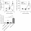
Fcγ RIII −/− mice display early inflammatory cell recruitment into the lungs, as compared with wild‐type (WT) mice. WT and Fcγ RIII −/− C57BL/6 mice were mock‐inoculated (HEp‐2 non‐infectious supernatants) or human respiratory syncytial virus (hRSV) infected (1 × 107 PFU). (a) Flow cytometry for CD11b+ GR‐1+ cells in bronchoalveolar lavage (BAL) was performed at different time‐points. Data are represented as the differences in the percentage of inflammatory cells, between hRSV‐infected and mock‐inoculated mice for each day analysed. WT mice: closed circles; Fcγ RIII −/− mice: open circles. Data are means ± SEM of two pooled independent experiments and were analysed by one‐way analysis of variance using Bonferroni's Multiple Comparison Test (*P < 0·05) (b) Quantitative real‐time PCR analyses of lung from hRSV‐infected mice. The graph shows number of RSV‐nucleoprotein RNA copies detected per ng of cDNA in lungs from WT (close circles) and Fcγ RIII −/− (open circle) mice. Data are means ± SEM of two independent experiments analysed by Student's t‐test (**P < 0·01). (c) Sera from WT and Fcγ RIII −/− mice were collected 6 days after infection with hRSV to assess specific antibodies against hRSV by ELISA. Data represent 1/20 dilution of the serum. A serum from uninfected WT mouse (Uninfected) was used as a negative control and anti‐F RS‐348 mouse antibody as a positive control. Data are means ± SEM of two independent experiments. Data were analysed by one‐way analysis of variance and Bonferroni's Multiple Comparison Test (***P < 0·0001, ns: non‐significant).

Similar articles

Cited by

References

    1. Hacking D, Hull J. Respiratory syncytial virus – viral biology and the host response. J Infect 2002; 45:18–24. - PubMed
    1. Collins PL, Graham BS. Viral and host factors in human respiratory syncytial virus pathogenesis. J Virol 2008; 82:2040–55. - PMC - PubMed
    1. Domachowske JB, Rosenberg HF. Respiratory syncytial virus infection: immune response, immunopathogenesis, and treatment. Clin Microbiol Rev 1999; 12:298–309. - PMC - PubMed
    1. Mejias A, Chavez‐Bueno S, Jafri HS, Ramilo O. Respiratory syncytial virus infections: old challenges and new opportunities. Pediatr Infect Dis J 2005; 24(11 Suppl):S189–96, discussion S96‐7. - PubMed
    1. Chang J, Braciale TJ. Respiratory syncytial virus infection suppresses lung CD8+ T‐cell effector activity and peripheral CD8+ T‐cell memory in the respiratory tract. Nat Med 2002; 8:54–60. - PubMed

MeSH terms

LinkOut - more resources