. 2015 Nov 24;6(37):39740-55.
doi: 10.18632/oncotarget.5469.

Overexpression of CHI3L1 is associated with chemoresistance and poor outcome of epithelial ovarian carcinoma

Affiliations

Overexpression of CHI3L1 is associated with chemoresistance and poor outcome of epithelial ovarian carcinoma

Ying-Cheng Chiang et al. Oncotarget. .

Abstract

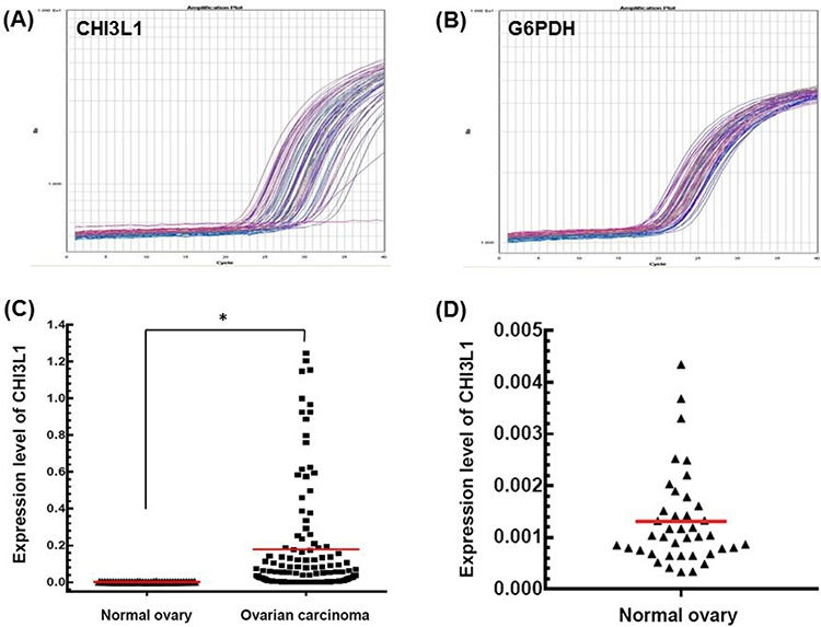
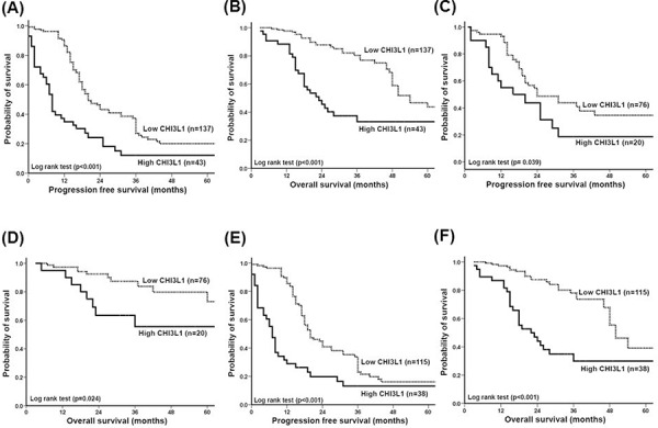
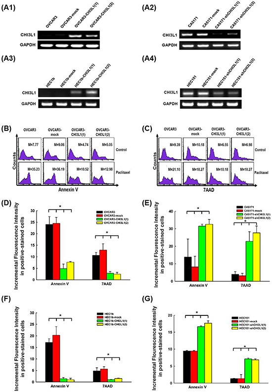
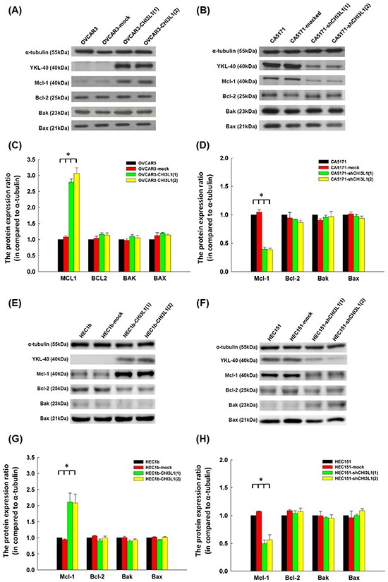
We propose CHI3L1 as a prognostic biomarker for patients with epithelial ovarian carcinoma (EOC) and also suggest possible biological functions of CHI3L1. We measured CHI3L1 expression with quantitative real time-polymerase chain reaction (qRT-PCR) in 180 women with EOC and evaluated correlations between CHI3L1 expression, clinicopathological characteristics, and the outcomes of the patients. The expression of CHI3L1 was higher in cancerous tissues than in normal tissues. The expression of CHI3L1 was also higher in patients with a serous histological type, advanced stage, and chemoresistance. Patients with high CHI3L1 expression had a shorter progression-free survival (p < 0.001)and overall survival (p < 0.001). Patients with high CHI3L1 expression also had a high risk of recurrence (p < 0.001)and death (p < 0.001). In vitro studies showed that CHI3L1 up-regulated the expression of anti-apoptotic Mcl-1 protein and hampered paclitaxel-induced apoptosis of ovarian cancer cells. These results suggest that CHI3L1 shows potential as a prognostic biomarker for EOC. CHI3L1 may promote chemoresistance via inhibition of drug-induced apoptosis by up-regulating Mcl-1.

Keywords: CHI3L1; apoptosis; chemoresistance; epithelial ovarian carcinoma.


Conflict of interest statement

CONFLICTS OF INTEREST

The authors declare no potential conflict of interest.

Figures

Figure 1
Figure 1. mRNA expression detected by quantitative real-time PCR
A. Representative figure of the quantification of CHI3L1 mRNA expression in tumor tissues. B. Representative figure of quantification of G6PDH mRNA expression in tumor tissues. C. CHI3L1 mRNA expression levels between normal and cancerous ovarian tissues. Black triangles indicated the normal ovarian tissues, and black squares indicated the ovarian cancer tissues.(*p < 0.05 by the Student's t-test) D. CHI3L1 mRNA expression levels of normal ovarian tissues were shown in detail.
Figure 2
Figure 2. Correlation of CHI3L1 expression with progression-free survival (PFS) and overall survival (OS) of patients with ovarian cancer
A. PFS of all 180 patients. Patients with a high CHI3L1 expression had a much shorter PFS (p < 0.001). B. OS of all 180 patients. Patients with a high CHI3L1 expression had a much, shorter OS (p < 0.001). C. PFS of the 96 patients whose residual tumor diameter was ≤ 1 cm. Patients with a high CHI3L1 expression had a shorter PFS (p = 0.039). D. OS of the 96 patients whose residual tumor diameter was ≤ 1 cm. Patients with a high CHI3L1 expression had a shorter OS (p = 0.024). E. PFS of the 153 patients who received adjuvant platinum-paclitaxel chemotherapy. Patients with a high CHI3L1 expression had a much shorter PFS (p < 0.001). F. OS of the 153 patients who received adjuvant platinum-paclitaxel chemotherapy. Patients with a high CHI3L1 expression had a much shorter OS (p < 0.001). All differences were calculated by the log rank test.
Figure 3
Figure 3. In vitro apoptotic assays of ovarian cancer (OVCAR3, CA5171) and endometrial cancer (HEC1b, HEC151) original cells, and their transfectants treated with respective cytotoxic drug
A. RT-PCR of CHI3L1 expression in the various cell lines. A1: The expression of CHI3L1 was higher in the OVCAR3-CHI3L1 transfectants than those in the original OVCAR3 and mock-transfectants. A2: The expression of CHI3L1 was lower in the CA5171-shCHI3L1 transfectants than those in the original CA5171 and mock-transfectants. A3: The expression of CHI3L1 was higher in the HEC1b-CHI3L1 transfectants than those in the original HEC1b and mock-transfectants. A4: The expression of CHI3L1 was lower in the HEC151-shCHI3L1 transfectants than those in the original HEC151 and mock-transfectants. B. Representative figures of flow cytometric analysis for annexin V-staining in original and various OVCAR3 transfectants treated with paclitaxel. C. Representative figures of flow cytometric analysis for 7AAD-stained in original and various OVCAR3 transfectants treated with paclitaxel. (The M values in (B) and (C) indicated the median value of fluorescence intensity of all cells stained with annexin V or 7AAD in original and various OVCAR3 transfectants). D. Bar figures of the incremental fluorescence intensity of Annexin V and 7AAD -positive cells in original and various OVCAR3 transfectants treated with paclitaxel for 48 hours (*p < 0.05 by ANOVA). The incremental fluorescence intensities of annexin V and 7AAD in the various OVCAR3 CHI3L1 transfectants were significantly lower than those in the original OVCAR3 and mock-transfected OVCAR3 cells. E. Bar figures of the incremental fluorescence intensity of Annexin V and 7AAD -positive cells in original and various CA5171 transfectants treated with paclitaxel for 48 hours (*p < 0.05 by ANOVA). The incremental fluorescence intensities of annexin V and 7AAD in the various CA5171 shCHI3L1 transfectants were higher than those in the original CA5171 and mock-transfected CA5171 cells. F. Bar figures of the incremental fluorescence intensity of Annexin V and 7AAD -positive cells in original and various HEC1b transfectants treated with paclitaxel for 48 hours (*p < 0.05 by ANOVA). The incremental fluorescence intensities of annexin V and 7AAD in the various HEC1b CHI3L1 transfectants were lower than those in the original HEC1b and mock-transfected HEC1b cells. G. Bar figures of the incremental fluorescence intensity of Annexin V and 7AAD -positive cells in original and various HEC151 transfectants treated with paclitaxel for 48 hours (*p < 0.05 by ANOVA). The incremental fluorescence intensities of annexin V and 7AAD in the various HEC151 shCHI3L1 transfectants were higher than those in the original HEC151 and mock-transfected HEC151 cells.
Figure 4
Figure 4. Western blot analysis of ovarian cancer (OVCAR3, CA5171) and endometrial cancer (HEC1b, HEC151) original cells and their transfectants
A. Representative figures of Western blots of apoptosis-related molecules in OVACR3 original cells and their transfectants. B. Representative figures of Western blots of apoptosis-related molecules in CA5171 original cells and their transfectants. C. Bar figures of protein expressions of various molecules in OVACR3 original cells and their transfectants. The expressions of Mcl-1 increased significantly in the CHI3L1-transfected OVCAR3 cells (*p < 0.05 by ANOVA). D. Bar figures of protein expressions of various molecules in CA5171 original cells and their transfectants. The expressions of Mcl-1 decreased significantly in the shCHI3L1-transfected CA5171 cells (*p < 0.05 by ANOVA). E. Representative figures of Western blots of apoptosis-related molecules in HEC1b original cells and their transfectants. F. Representative figures of Western blots of apoptosis-related molecules in HEC151 original cells and their transfectants. G. Bar figures of protein expressions of various molecules in HEC1b original cells and their transfectants. The expressions of Mcl-1 increased significantly in the CHI3L1-transfected HEC1b cells (*p < 0.05 by ANOVA). H. Bar figures of protein expressions of various molecules in HEC151 original cells and their transfectants. The expressions of Mcl-1 decreased significantly in the shCHI3L1-transfected HEC151 cells (*p < 0.05 by ANOVA).

References

    1. Siegel R, Naishadham D, Jemal A. Cancer statistics, 2012. CA Cancer J Clin. 2012;62:10–29. - PubMed
    1. Chiang YC, Chen CA, Chiang CJ, Hsu TH, Lin MC, You SL, Cheng WF, Lai MS. Trends in incidence and survival outcome of epithelial ovarian cancer: 30-year national population-based registry in Taiwan. J Gynecol Oncol. 2013;24:342–351. - PMC - PubMed
    1. Agarwal R, Kaye SB. Ovarian cancer: strategies for overcoming resistance to chemotherapy. Nat Rev Cancer. 2003;3:502–516. - PubMed
    1. Stuart GC. First-line treatment regimens and the role of consolidation therapy in advanced ovarian cancer. Gynecologic Oncology. 2003;90:S8–15. - PubMed
    1. O'Gorman DM, Cotter TG. Molecular signals in anti-apoptotic survival pathways. Leukemia. 2001;15:21–34. - PubMed

Publication types

MeSH terms