Beta Cell Mass Restoration in Alloxan-Diabetic Mice Treated with EGF and Gastrin
- PMID: 26452142
- PMCID: PMC4599944
- DOI: 10.1371/journal.pone.0140148
Beta Cell Mass Restoration in Alloxan-Diabetic Mice Treated with EGF and Gastrin
Abstract
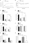
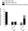
One week of treatment with EGF and gastrin (EGF/G) was shown to restore normoglycemia and to induce islet regeneration in mice treated with the diabetogenic agent alloxan. The mechanisms underlying this regeneration are not fully understood. We performed genetic lineage tracing experiments to evaluate the contribution of beta cell neogenesis in this model. One day after alloxan administration, mice received EGF/G treatment for one week. The treatment could not prevent the initial alloxan-induced beta cell mass destruction, however it did reverse glycemia to control levels within one day, suggesting improved peripheral glucose uptake. In vitro experiments with C2C12 cell line showed that EGF could stimulate glucose uptake with an efficacy comparable to that of insulin. Subsequently, EGF/G treatment stimulated a 3-fold increase in beta cell mass, which was partially driven by neogenesis and beta cell proliferation as assessed by beta cell lineage tracing and BrdU-labeling experiments, respectively. Acinar cell lineage tracing failed to show an important contribution of acinar cells to the newly formed beta cells. No appearance of transitional cells co-expressing insulin and glucagon, a hallmark for alpha-to-beta cell conversion, was found, suggesting that alpha cells did not significantly contribute to the regeneration. An important fraction of the beta cells significantly lost insulin positivity after alloxan administration, which was restored to normal after one week of EGF/G treatment. Alloxan-only mice showed more pronounced beta cell neogenesis and proliferation, even though beta cell mass remained significantly depleted, suggesting ongoing beta cell death in that group. After one week, macrophage infiltration was significantly reduced in EGF/G-treated group compared to the alloxan-only group. Our results suggest that EGF/G-induced beta cell regeneration in alloxan-diabetic mice is driven by beta cell neogenesis, proliferation and recovery of insulin. The glucose-lowering effect of the treatment might play an important role in the regeneration process.
Conflict of interest statement
Figures


Similar articles
-
A combination of cytokines EGF and CNTF protects the functional beta cell mass in mice with short-term hyperglycaemia.Diabetologia. 2016 Sep;59(9):1948-58. doi: 10.1007/s00125-016-4023-3. Epub 2016 Jun 18. Diabetologia. 2016. PMID: 27318836
-
Combined gastrin and epidermal growth factor treatment induces islet regeneration and restores normoglycaemia in C57Bl6/J mice treated with alloxan.Diabetologia. 2004 Feb;47(2):259-65. doi: 10.1007/s00125-003-1287-1. Epub 2003 Dec 10. Diabetologia. 2004. PMID: 14666367
-
Liraglutide improves pancreatic Beta cell mass and function in alloxan-induced diabetic mice.PLoS One. 2015 May 4;10(5):e0126003. doi: 10.1371/journal.pone.0126003. eCollection 2015. PLoS One. 2015. PMID: 25938469 Free PMC article.
-
Pharmacological treatment of chronic diabetes by stimulating pancreatic beta-cell regeneration with systemic co-administration of EGF and gastrin.Pharmacol Toxicol. 2002 Dec;91(6):414-20. doi: 10.1034/j.1600-0773.2002.910621.x. Pharmacol Toxicol. 2002. PMID: 12688387 Review.
-
In vivo regeneration of insulin-producing beta-cells.Adv Exp Med Biol. 2010;654:627-40. doi: 10.1007/978-90-481-3271-3_27. Adv Exp Med Biol. 2010. PMID: 20217517 Review.
Cited by
-
Coordinated interactions between endothelial cells and macrophages in the islet microenvironment promote β cell regeneration.NPJ Regen Med. 2021 Apr 6;6(1):22. doi: 10.1038/s41536-021-00129-z. NPJ Regen Med. 2021. PMID: 33824346 Free PMC article.
-
A combination of cytokines EGF and CNTF protects the functional beta cell mass in mice with short-term hyperglycaemia.Diabetologia. 2016 Sep;59(9):1948-58. doi: 10.1007/s00125-016-4023-3. Epub 2016 Jun 18. Diabetologia. 2016. PMID: 27318836
-
A Supportive Role of Mesenchymal Stem Cells on Insulin-Producing Langerhans Islets with a Specific Emphasis on The Secretome.Biomedicines. 2023 Sep 18;11(9):2558. doi: 10.3390/biomedicines11092558. Biomedicines. 2023. PMID: 37761001 Free PMC article. Review.
-
Pancreatic Progenitors: There and Back Again.Trends Endocrinol Metab. 2019 Jan;30(1):4-11. doi: 10.1016/j.tem.2018.10.002. Epub 2018 Nov 28. Trends Endocrinol Metab. 2019. PMID: 30502039 Free PMC article. Review.
-
Islet Regeneration and Pancreatic Duct Glands in Human and Experimental Diabetes.Front Cell Dev Biol. 2022 Feb 4;10:814165. doi: 10.3389/fcell.2022.814165. eCollection 2022. Front Cell Dev Biol. 2022. PMID: 35186929 Free PMC article.
References
-
- Song I, Muller C, Louw J, Bouwens L. Regulating the Beta Cell Mass as a Strategy for Type-2 Diabetes Treatment. Curr Drug Targets. 2015. Epub 2015/02/06. CDT-EPUB-64988 [pii]. . - PubMed
Publication types
MeSH terms
Substances
LinkOut - more resources
Full Text Sources
Other Literature Sources
Research Materials

