

Comment
doi: 10.7554/eLife.11409.
Mapping neural circuits with CLARITY
Affiliations
- PMID: 26452201
- PMCID: PMC4598891
- DOI: 10.7554/eLife.11409
Item in Clipboard
Comment
Mapping neural circuits with CLARITY
Elife.
.
Abstract
The use of whole-brain imaging has shed new light on the organization of the dopamine system.
Keywords: anatomy; dopamine; input; monosynaptic; mouse; neuroscience; rabies virus; striatum.
Conflict of interest statement
Figures
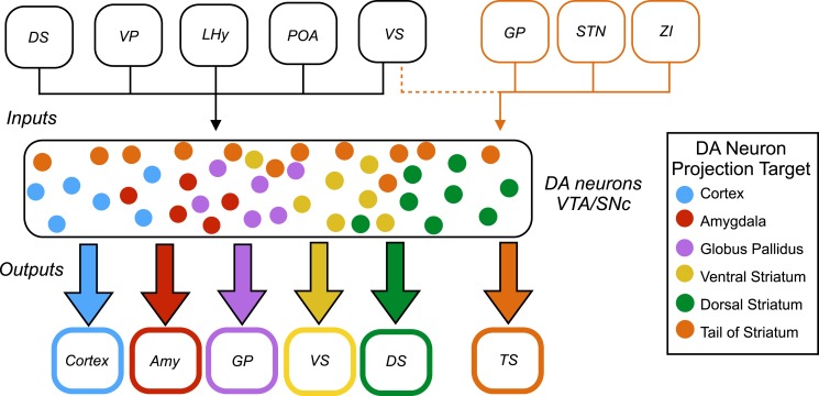
Dopamine (DA) neurons are found in the ventral tegmental area (VTA) and the substantia nigra pars compacta (SNc). Dopamine neurons projecting to the tail of the striatum (TS) receive the majority of their input from the globus pallidus (GP), the subthalamic nucleus (STN), and the zona incerta (ZI), with a small amount of input coming from the ventral striatum (VS). Dopamine neurons that project to the cortex, amygdala (Amy), globus pallidus, ventral striatum and dorsal striatum (DS) receive the majority of their inputs from the following regions: the ventral striatum, dorsal striatum, ventral pallidum (VP), lateral hypothalamus (LHy), and preoptic area (POA). In Parkinson’s disease, dopamine neurons projecting to the posterior putamen (which is functionally similar to the tail of the striatum) are the first to degenerate (Kish et al., 1988), so their unique pattern of inputs is especially interesting to researchers.
Comment on
-
Dopamine neurons projecting to the posterior striatum form an anatomically distinct subclass.Elife. 2015 Aug 31;4:e10032. doi: 10.7554/eLife.10032. Elife. 2015. PMID: 26322384 Free PMC article.
References
-
- Chung K, Wallace J, Kim SY, Kalyanasundaram S, Andalman AS, Davidson TJ, Mirzabekov JJ, Zalocusky KA, Mattis J, Denisin AK, Pak S, Bernstein H, Ramakrishnan C, Grosenick L, Gradinaru V, Deisseroth K. Structural and molecular interrogation of intact biological systems. Nature. 2013;497:332–337. doi: 10.1038/nature12107. - DOI - PMC - PubMed
Publication types
MeSH terms
Grants and funding
LinkOut - more resources
Full Text Sources
