. 2015 Oct 13;6(31):32154-60.
doi: 10.18632/oncotarget.5168.

Hypoxia-induced endoplasmic reticulum stress characterizes a necrotic phenotype of pancreatic cancer

Affiliations

Hypoxia-induced endoplasmic reticulum stress characterizes a necrotic phenotype of pancreatic cancer

Bo Kong et al. Oncotarget. .

Abstract

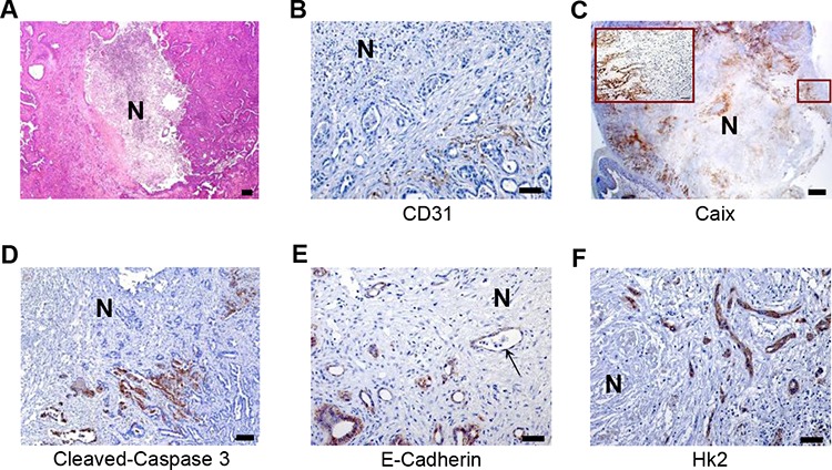
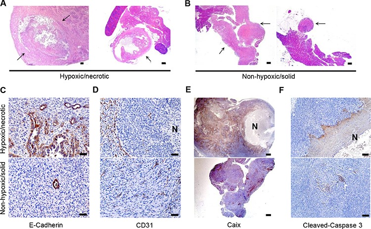
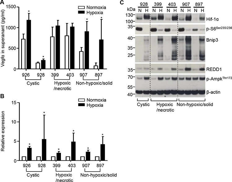
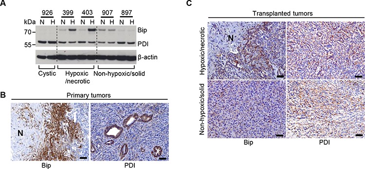
Stromal fibrosis and tissue necrosis are major histological sequelae of hypoxia. The hypoxia-to-fibrosis sequence is well-documented in pancreatic ductal adenocarcinoma (PDAC). However, hypoxic and necrotic PDAC phenotypes are insufficiently characterized. Recently, reduction of tuberous sclerosis expression in mice together with oncogenic Kras demonstrated a rapidly metastasizing phenotype with histologically eccentric necrosis, transitional hypoxia and devascularisation. We established cell lines from these tumors and transplanted them orthotopically into wild-type mice to test their abilities to recapitulate the histological features of the primary lesions. Notably, the necrotic phenotype was reproduced by only a subset of cell lines while others gave rise to dedifferentiated tumors with significantly reduced necrosis. In vitro analysis of the necrotic tumor-inducing cell lines revealed that these cells released a significant amount of vascular endothelial growth factor A (Vegfa). However, its release was not further increased under hypoxic conditions. Defective hypoxia-induced Vegfa secretion was not due to impaired Vegfa transcription or hypoxia-inducible factor 1-alpha activation, but rather a result of hypoxia-induced endoplasmic reticulum (ER) stress. We thus identified hypoxia-induced ER stress as an important pathway in PDACs with tissue necrosis and rapid metastasis.

Keywords: ER stress; VEGFA; hypoxia; necrosis; pancreatic cancer.


Conflict of interest statement

CONFLICTS OF INTEREST

None.

Figures

Figure 1
Figure 1. Identification of a hypoxic/necrotic tumor phenotype
A. Representative H&E-stained sections of necrotic PDACs; scale bar: 100 μm, B–D. IHC studies of CD31, Caix and cleaved-caspase 3 as surrogate markers for vessel localization (scale bar: 50 μm), hypoxic regions (scale bar: 500 μm) and apoptotic cells (scale bar: 100 μm) in metastatic PDACs. E–F. IHC studies for E-Cadherin and Hk2 show loss of membrane staining of E-Cadherin (arrow) and increased expression of Hk2 in cancer cells in the vicinity of tissue necrosis (also hypoxic regions) in metastatic PDACs, scale bar: 50 μm, N: necrosis.
Figure 2
Figure 2. In vivo screening of PDAC cells with an in vivo preserved hypoxic/necrotic phenotype
A–B. H&E–stained sections show centrally-necrotic tumors formed after orthotopic implantation of the hypoxic/necrotic cells and more solid forms of tumors formed by dedifferentiated cells, scale bar: 500 μm; arrow: tumor tissues; C. IHC with anti-E-Cadherin demonstrates a high proportion of anaplastic components in tumors developing from the hypoxic/necrotic cells, but not in tumors developing from the dedifferentiated cells, scale bar: 50 μm; D–F. IHC of anti-CD31 (scale bar: 50 μm), anti-Caix (scale bar: 500 μm) and anti-cleaved-caspase 3 (scale bar: 100 μm) show devascularized tumor regions, hypoxic zones and apoptosis in the necrotic tumors forming after transplantation of the hypoxic/necrotic cells, but not in the anaplastic tumors forming after transplantation of the dedifferentiated cells; N: necrosis.
Figure 3
Figure 3. Compromised hypoxia-driven VEGFA secretion in PDAC cells with the hypoxic/necrotic phenotype
A. ELISA assays show the release of VEGFA into the supernatants of hypoxic/necrotic cells (399 and 403 cells), dedifferentiated cells (907 and 897 cells) and “cystic” cells (926 and 928 cells) after exposure to hypoxia for 24 hours. Values shown are obtained from at least three independent experiments; *p < 0.05; B. VEGFA mRNA levels (measured by QRT-PCR) after induction of hypoxia in all tested cell lines. Data are presented as relative expression (normalized to the median of the respective expression level under normoxia); an unpaired t-test was performed; values shown are obtained from at least three independent experiments; *p < 0.05; C, Western-blot analysis shows expression of Hif1alpha, p-S6Ser235/236, Bnip3, Redd1 and p-AmpkThr172 in hypoxic/necrotic cells (399 and 403 cells), dedifferentiated cells (907 and 897 cells) and “cystic” cells (928 cells) after exposure to hypoxia for 24 hours; N: normoxia, H: hypoxia; one representative blot out of three independent experiments is shown.
Figure 4
Figure 4. Sensitivity of the hypoxic/necrotic cells to hypoxia-induced ER stress
A. Western-blot analysis demonstrates induced expression of Bip and PDI in hypoxic/necrotic cells (399, 403), in dedifferentiated cells (907, 897) and in cystic cells (926) after exposure to hypoxia for 24 hours; N: normoxia, H: hypoxia; one representative blot out of three independent experiments is shown, B. Representative IHC pictures show expression of Bip and PDI in metastatic PDACs, scale bar: 50 μm, C. Representative IHC pictures show expression of Bip and PDI in the transplanted tumor tissues induced by transplantation of hypoxic/necrotic or dedifferentiated cells, scale bar: 50 μm.

References

    1. Koong AC, Mehta VK, Le QT, Fisher GA, Terris DJ, Brown JM, Bastidas AJ, Vierra M. Pancreatic tumors show high levels of hypoxia. International journal of radiation oncology, biology, physics. 2000;48:919–922. - PubMed
    1. Siegel R, Naishadham D, Jemal A. Cancer statistics, 2013. CA Cancer J Clin. 2013;63:11–30. - PubMed
    1. Kitada T, Seki S, Sakaguchi H, Sawada T, Hirakawa K, Wakasa K. Clinicopathological significance of hypoxia-inducible factor-1alpha expression in human pancreatic carcinoma. Histopathology. 2003;43:550–555. - PubMed
    1. Shibaji T, Nagao M, Ikeda N, Kanehiro H, Hisanaga M, Ko S, Fukumoto A, Nakajima Y. Prognostic significance of HIF-1 alpha overexpression in human pancreatic cancer. Anticancer Res. 2003;23:4721–4727. - PubMed
    1. Hoffmann AC, Mori R, Vallbohmer D, Brabender J, Klein E, Drebber U, Baldus SE, Cooc J, Azuma M, Metzger R, Hoelscher AH, Danenberg KD, Prenzel KL, Danenberg PV. High expression of HIF1a is a predictor of clinical outcome in patients with pancreatic ductal adenocarcinomas and correlated to PDGFA, VEGF, and bFGF. Neoplasia. 2008;10:674–679. - PMC - PubMed

Publication types

Substances