Pulmonary Kaposi sarcoma and disseminated Mycobacterium genavense infection in an HIV-infected patient
- PMID: 26452414
- PMCID: PMC4600798
- DOI: 10.1136/bcr-2015-211683
Pulmonary Kaposi sarcoma and disseminated Mycobacterium genavense infection in an HIV-infected patient
Abstract
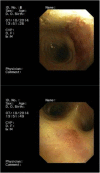
We report a case of Kaposi sarcoma (KS) and disseminated infection by Mycobacterium genavense in a 40-year-old HIV-positive man with CD4+ T-cell count 5/µL. He presented with anorexia, diarrhoea, cachexia and multiple firm violaceous nodules distributed over the face, neck and upper and lower extremities. Biopsy of a skin nodule was performed, confirming KS. Immunoperoxidase staining for human herpesvirus 8 was strongly positive. Endoscopic examination revealed erosive duodenopathy. Multiple biopsy samples showed numerous acid-fast bacilli at direct microscopic examination. Real-time PCR (RT-PCR) identified M. genavense. A CT scan showed diffuse pulmonary infiltrates with a 'tree-in-bud' appearance, striking splenomegaly and abdominal lymphadenopathy. A bronchoscopy was performed, revealing typical Kaposi's lesions in the upper respiratory tract. RT-PCR of bronchial aspirate identified M. genavense and Pneumocystis jirovecii. Despite treatment with highly active antiretroviral therapy, antimycobacterial therapy and trimethoprim/sulfamethoxazole, the outcome was fatal.
2015 BMJ Publishing Group Ltd.
Figures


Similar articles
-
Mycobacterium genavense infections: a retrospective multicenter study in France, 1996-2007.Medicine (Baltimore). 2011 Jul;90(4):223-230. doi: 10.1097/MD.0b013e318225ab89. Medicine (Baltimore). 2011. PMID: 21694645
-
Chronic cough conundrum: a case report of a new diagnosis of HIV and pulmonary Kaposi's sarcoma.BMC Pulm Med. 2017 Mar 20;17(1):52. doi: 10.1186/s12890-017-0395-5. BMC Pulm Med. 2017. PMID: 28320359 Free PMC article.
-
Pulmonary Kaposi's Sarcoma - Initial Presentation of HIV Infection.Folia Med (Plovdiv). 2019 Dec 31;61(4):643-649. doi: 10.3897/folmed.61.e47945. Folia Med (Plovdiv). 2019. PMID: 32337885
-
Disseminated mycobacterium genavense infection with central nervous system involvement in an HIV patient: a case report and literature review.BMC Infect Dis. 2024 Apr 24;24(1):437. doi: 10.1186/s12879-024-09316-x. BMC Infect Dis. 2024. PMID: 38658840 Free PMC article. Review.
-
Mycobacterium genavense infection presenting as a solitary brain mass in a patient with AIDS: case report and review.Clin Infect Dis. 1994 Dec;19(6):1152-4. doi: 10.1093/clinids/19.6.1152. Clin Infect Dis. 1994. PMID: 7888551 Review.
Cited by
-
Disseminated infection by Mycobacterium genavense in an HIV-1 infected patient.IDCases. 2020 Jul 27;21:e00926. doi: 10.1016/j.idcr.2020.e00926. eCollection 2020. IDCases. 2020. PMID: 32775210 Free PMC article.
-
An unusual case of abdominal mycobacterial infection: Case report and literature review.South Afr J HIV Med. 2019 Aug 28;20(1):993. doi: 10.4102/sajhivmed.v20i1.993. eCollection 2019. South Afr J HIV Med. 2019. PMID: 31534791 Free PMC article.
References
-
- Ferla LL, Pinzone MR, Nunnari G et al. . Kaposi's sarcoma in HIV-positive patients: the state of art in the HAART-era. Eur Rev Med Pharmacol Sci 2013;17:2354–65. - PubMed
-
- Dezube BJ, Pantanowitz L, Aboulafia DM. Management of AIDS-related Kaposi sarcoma: advances in target discovery and treatment. AIDS Read 2004;14:236–8, 243–4, 251–3. - PubMed
Publication types
MeSH terms
Substances
LinkOut - more resources
Full Text Sources
Other Literature Sources
Medical
Research Materials
