

Contrast-enhanced ultrasonography in Takayasu arteritis: watching and monitoring the arterial inflammation
- PMID: 26452525
- PMCID: PMC4612304
- DOI: 10.1136/bcr-2015-211094
Contrast-enhanced ultrasonography in Takayasu arteritis: watching and monitoring the arterial inflammation
Abstract
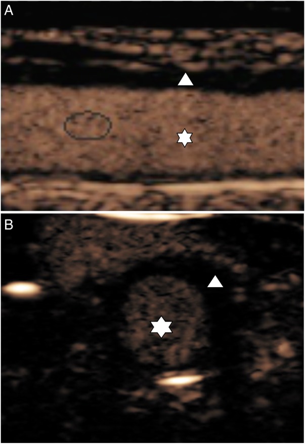
A 43-year-old man was diagnosed with Takayasu arteritis, and treated with methotrexate and corticosteroids. While under treatment and with normal biological inflammatory parameters, he experienced an ischaemic stroke, successfully treated with intravenous thrombolysis (alteplase). The B-mode ultrasound examination revealed circumferential wall thickening of the left common carotid artery. Contrast-enhanced ultrasonography showed a progressive arterial wall enhancement of the left common carotid artery. This pathological enhancement indicates neovascularisation of the arterial wall, which is supposed to correlate with active vascular inflammation. After an increase in immunosuppressive treatment, follow-up contrast-enhanced ultrasonography no longer showed artery wall enhancement. Contrast-enhanced ultrasound examination is an inexpensive, reproducible and minimally invasive method, providing dynamic information on arterial wall neovascularisation and thus inflammation. This case illustrates that contrast-enhanced ultrasonography can be a useful tool for the management and follow-up of Takayasu arteritis, and its use as a marker of disease activity and arterial inflammation in Takayasu arteritis should be evaluated in further studies.
2015 BMJ Publishing Group Ltd.
Figures

References
Publication types
MeSH terms
Substances
LinkOut - more resources
Full Text Sources
Other Literature Sources
Medical