. 2015 Dec 1;309(11):H1837-45.
doi: 10.1152/ajpheart.00463.2015. Epub 2015 Oct 9.

Purinergic glio-endothelial coupling during neuronal activity: role of P2Y1 receptors and eNOS in functional hyperemia in the mouse somatosensory cortex

Affiliations

Purinergic glio-endothelial coupling during neuronal activity: role of P2Y1 receptors and eNOS in functional hyperemia in the mouse somatosensory cortex

Peter Toth et al. Am J Physiol Heart Circ Physiol. .

Abstract

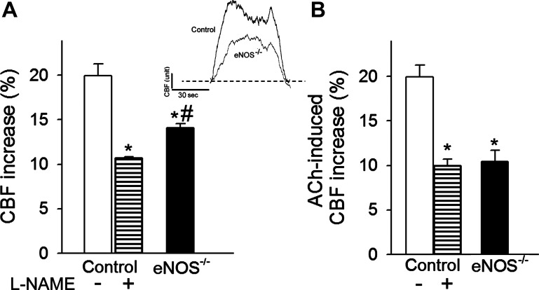
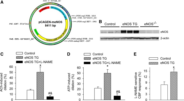
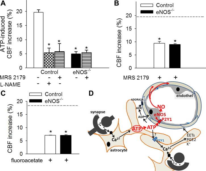
Impairment of moment-to-moment adjustment of cerebral blood flow (CBF) via neurovascular coupling is thought to play a critical role in the genesis of cognitive impairment associated with aging and pathological conditions associated with accelerated cerebromicrovascular aging (e.g., hypertension, obesity). Although previous studies demonstrate that endothelial dysfunction plays a critical role in neurovascular uncoupling in these conditions, the role of endothelial NO mediation in neurovascular coupling responses is not well understood. To establish the link between endothelial function and functional hyperemia, neurovascular coupling responses were studied in mutant mice overexpressing or deficient in endothelial NO synthase (eNOS), and the role of P2Y1 receptors in purinergic glioendothelial coupling was assessed. We found that genetic depletion of eNOS (eNOS(-/-)) and pharmacological inhibition of NO synthesis significantly decreased the CBF responses in the somatosensory cortex evoked by whisker stimulation and by administration of ATP. Overexpression of eNOS enhanced NO mediation of functional hyperemia. In control mice, the selective and potent P2Y1 receptor antagonist MRS2179 attenuated both whisker stimulation-induced and ATP-mediated CBF responses, whereas, in eNOS(-/-) mice, the inhibitory effects of MRS2179 were blunted. Collectively, our findings provide additional evidence for purinergic glio-endothelial coupling during neuronal activity, highlighting the role of ATP-mediated activation of eNOS via P2Y1 receptors in functional hyperemia.

Keywords: astrocyte; dementia; endothelial dysfunction; endothelial nitric oxide synthase; endothelium; vascular cognitive impairment.


Figures

Fig. 1.
Fig. 1.
Endothelial nitric oxide synthase (eNOS) contributes to neurovascular coupling. A: increases in cerebral blood flow (CBF; expressed as percentage of baseline) measured above the barrel field of the primary somatosensory cortex in response to whisker stimulation in control mice in the presence and absence of the nitric oxide synthase inhibitor Nω-nitro-l-arginine methyl ester (l-NAME) and in mice with genetic depletion of eNOS (eNOS−/−). Data are means ± SE (n = 8, *P < 0.05 vs. control, #P < 0.5 vs. control + l-NAME). Inset: representative traces of CBF measured with a laser Doppler probe above the whisker barrel cortex during contralateral whisker stimulation (5 Hz) in control and eNOS−/− mice. 1 AU corresponds to ∼5% increase in CBF from baseline. B: CBF responses elicited by topical administration of acetylcholine (ACh, 10−5 mol/l) to the barrel field of control and eNOS−/− mice (n = 5 in each group, *P < 0.05 vs. control). Data are means ± SE.
Fig. 2.
Fig. 2.
eNOS overexpression increases the NO-mediated part of neurovascular coupling response. A: construct used to develop the transgenic (TG) mouse overexpressing eNOS (see methods). B: representative Western blot image showing expression of eNOS in the cortex of control wild-type mice, eNOS TG mice, and eNOS−/− mice. C and D: overexpression of eNOS (eNOS TG) is associated with significantly enhanced dilation of isolated cerebral vessels to ACh (10−7 mol/l; C) and ATP (10−7 mol/l; D), which are significantly decreased by the NOS inhibitor l-NAME (10−4 mol/l for 30 min). Data are means ± SE. (n = 5 in each group, *P < 0.05 eNOS TG vs. control, #P < 0.05 eNOS TG + l-NAME vs. eNOS TG; &P < 0.05 eNOS TG + l-NAME vs. control). E: overexpression of eNOS significantly enhances the l-NAME-sensitive, NO-mediated portion of the CBF response (calculated based on the percentage decline in CBF in the presence of l-NAME) measured above the barrel field of the primary somatosensory cortex in response to whisker stimulation. Data are means ± SE (n = 5, *P < 0.05 eNOS TG vs. control).
Fig. 3.
Fig. 3.
ATP-mediated CBF responses, role of P2Y1 receptors in NO mediation of neurovascular coupling. A: increases in CBF (expressed as a percentage of baseline) in the barrel field of the primary somatosensory cortex of control and eNOS−/− mice in response to topical administration of ATP in the presence and absence of the potent, selective, competitive P2Y1 receptor antagonist MRS2179 (6 × 10⁻⁵ mol/l for 20 min) or the NOS inhibitor l-NAME (10−4 mol/l for 20 min) (n = 6, *P < 0.05 vs. control). CBF was measured with a laser Doppler probe above the whisker barrel cortex. B and C: increases in CBF in the barrel field of the primary somatosensory cortex of control and eNOS−/− mice induced by contralateral whisker stimulation (5 Hz) in the presence of MRS2179 (6 × 10⁻⁵ mol/l for 20 min; B) or the glial-specific metabolic toxin fluoroacetate (3 × 10⁻⁴ mol/l for 20 min, C). Dashed line indicates the mean of the control responses (n = 6, *P < 0.05 vs. control). Data are means ± SE. D: scheme depicting purinergic glio-endothelial coupling during neuronal activity, highlighting the role of ATP-mediated activation of eNOS via P2Y1 receptors in functional hyperemia. The model acknowledges that multiple parallel astrocytic pathways exist [including synthesis of epoxyeicosatrienoic acids (EETs), prostaglandins (PGE2), and release of K+, which hyperpolarizes vascular smooth muscle cells] that are activated simultaneously to regulate blood flow and that ATP and its hydrolysis products [ADP and adenosine (ADE)] can evoke arteriolar dilation via adenosine A2 receptors located on the smooth muscle cells, as well.

References

    1. Aldasoro M, Martinez C, Vila JM, Medina P, Lluch S. Influence of endothelial nitric oxide on adrenergic contractile responses of human cerebral arteries. J Cereb Blood Flow Metab 16: 623–628, 1996. - PubMed
    1. Arcuino G, Lin JH, Takano T, Liu C, Jiang L, Gao Q, Kang J, Nedergaard M. Intercellular calcium signaling mediated by point-source burst release of ATP. Proc Natl Acad Sci USA 99: 9840–9845, 2002. - PMC - PubMed
    1. Austin SA, Santhanam AV, Hinton DJ, Choi DS, Katusic ZS. Endothelial nitric oxide deficiency promotes Alzheimer's disease pathology. J Neurochem 127: 691–700, 2013. - PMC - PubMed
    1. Ayata C, Ma J, Meng W, Huang P, Moskowitz MA. l-NA-sensitive rCBF augmentation during vibrissal stimulation in type III nitric oxide synthase mutant mice. J Cereb Blood Flow Metab 16: 539–541, 1996. - PubMed
    1. Bannerman DM, Chapman PF, Kelly PA, Butcher SP, Morris RG. Inhibition of nitric oxide synthase does not impair spatial learning. J Neurosci 14: 7404–7414, 1994. - PMC - PubMed

Publication types

MeSH terms

LinkOut - more resources