

Skeletal muscle as an endocrine organ: PGC-1α, myokines and exercise
- PMID: 26453501
- PMCID: PMC4657151
- DOI: 10.1016/j.bone.2015.02.008
Skeletal muscle as an endocrine organ: PGC-1α, myokines and exercise
Abstract
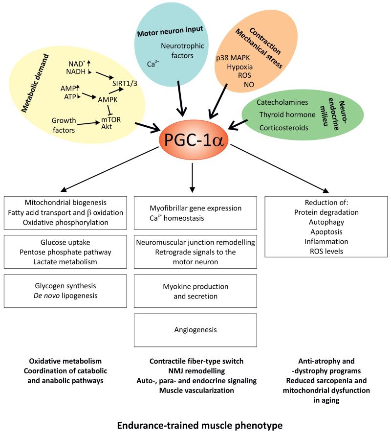
An active lifestyle is crucial to maintain health into old age; inversely, sedentariness has been linked to an elevated risk for many chronic diseases. The discovery of myokines, hormones produced by skeletal muscle tissue, suggests the possibility that these might be molecular mediators of the whole body effects of exercise originating from contracting muscle fibers. Even though less is known about the sedentary state, the lack of contraction-induced myokines or the production of a distinct set of hormones in the inactive muscle could likewise contribute to pathological consequences in this context. In this review, we try to summarize the most recent developments in the study of muscle as an endocrine organ and speculate about the potential impact on our understanding of exercise and sedentary physiology, respectively. This article is part of a Special Issue entitled "Muscle Bone Interactions".
Keywords: Exercise; Inflammation; Myokines; PGC-1α; Skeletal muscle.
Copyright © 2015 Elsevier Inc. All rights reserved.
Figures

References
-
- Hoppeler H, et al. Molecular mechanisms of muscle plasticity with exercise. Comprehensive Physiology. 2011;1(3):1383–412. - PubMed
-
- Schiaffino S, Reggiani C. Fiber types in mammalian skeletal muscles. Physiological reviews. 2011;91(4):1447–531. - PubMed
-
- Blaauw B, Schiaffino S, Reggiani C. Mechanisms modulating skeletal muscle phenotype. Comprehensive Physiology. 2013;3(4):1645–87. - PubMed
-
- Nikolaidis MG, et al. Redox biology of exercise: an integrative and comparative consideration of some overlooked issues. The Journal of experimental biology. 2012;215(Pt 10):1615–25. - PubMed
Publication types
MeSH terms
Substances
Grants and funding
LinkOut - more resources
Full Text Sources
Other Literature Sources
Medical
