. 2016 Jun;27(6):1689-700.
doi: 10.1681/ASN.2015040415. Epub 2015 Oct 9.

Interaction of the EGF Receptor and the Hippo Pathway in the Diabetic Kidney

Affiliations

Interaction of the EGF Receptor and the Hippo Pathway in the Diabetic Kidney

Jianchun Chen et al. J Am Soc Nephrol. 2016 Jun.

Abstract

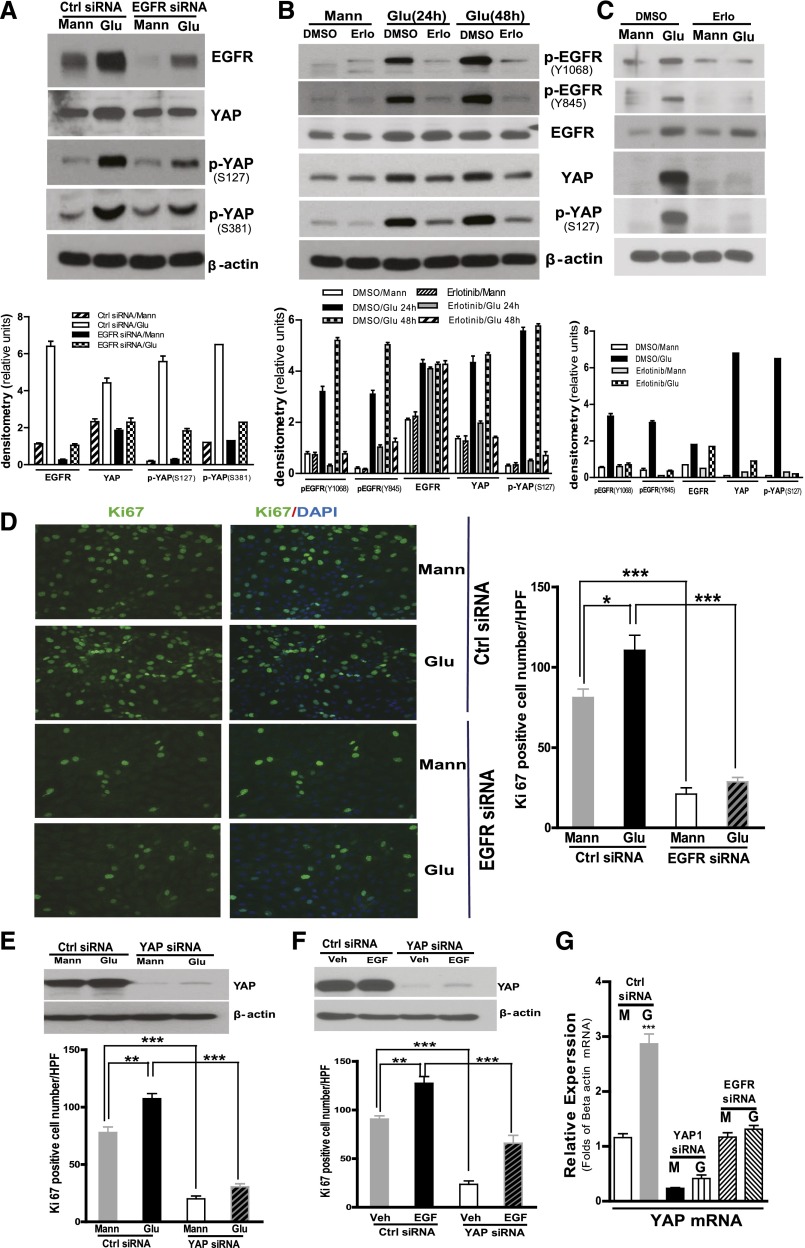
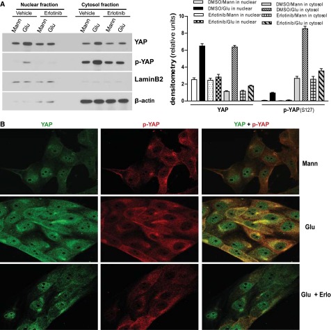
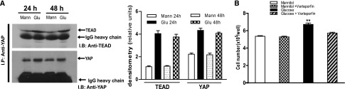
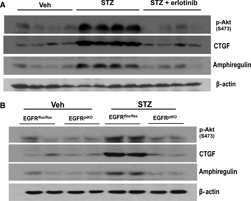
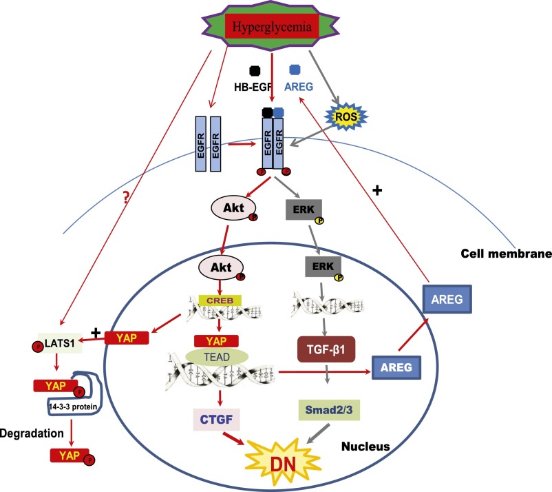
Activation of the EGF receptor (EGFR) or the Hippo signaling pathway can control cell proliferation, apoptosis, and differentiation, and the dysregulation of these pathways can contribute to tumorigenesis. Previous studies showed that activation of EGFR signaling in renal epithelial cells can exacerbate diabetic kidney injury. Moreover, EGFR has been implicated in regulating the Hippo signaling pathway in Drosophila; thus, we examined this potential interaction in mammalian diabetic kidney disease. Yes-associated protein (YAP) is a transcriptional regulator regulated by the Hippo signaling pathway. We found YAP protein expression and phosphorylation were upregulated in diabetic mouse renal proximal tubule epithelial cells, which were inhibited in diabetic proximal tubule EGFR-knockout mice (EGFR(ptKO)) or administration of an EGFR tyrosine kinase inhibitor erlotinib. Furthermore, activation of an EGFR-PI3K-Akt-CREB signaling pathway mediated YAP gene expression and YAP nuclear translocation and interaction with the TEA domain (TEAD) transcription factor complex, which led to upregulated expression of two TEAD-dependent genes, the connective tissue growth factor and amphiregulin genes. In a renal proximal tubule cell line, either pharmacologic or genetic inhibition of EGFR, Akt, or CREB blunted YAP expression in response to high-glucose treatment. Additionally, knocking down YAP expression by specific siRNA inhibited cell proliferation in response to high glucose or exogenous EGF. Therefore, these results link the Hippo pathway to EGFR-mediated renal epithelial injury in diabetes.

Keywords: cell signaling; diabetic nephropathy; epidermal growth factor.


Figures

Figure 1.
Figure 1.
EGFR dependence of increased YAP expression and phosphorylation in diabetic mouse proximal tubule epithelial cells. Wild-type balb/c mice 9–10 weeks old were subjected to five consecutive STZ injections followed by administration or no administration of erlotinib by gavage (80 mg/kg per day). The mice were euthanized at 2 weeks after development of hyperglycemia. Renal cortical tissues were collected and analyzed by immunoblotting (A) or immunofluorescence (B, Red:YAP; Green:LTA; Purple: DAPI) Original magnification, upper panel ×400; lower panel, ×1200. (C) EGFRptKO mice 9–10 weeks old and age-matched controls were made diabetic with STZ and euthanized at 2 weeks after development of hyperglycemia. Renal cortical tissue lysates were analyzed as in part A. (D) Renal cortical tissue lysates of diabetic db/db mice (at 24 weeks age) were analyzed by immunoblotting. n=5–7 mice/group. Shown are representative data from three separate experiments. Ctrl, control; Veh, vehicle.
Figure 2.
Figure 2.
Blocking EGFR expression or activation inhibited YAP protein expression and phosphorylation in cultured renal proximal tubule epithelial cells. (A) LLCPK-Cl4 cells were exposed to high glucose (Glu) for 24 hours after 3 days of control siRNA or EGFR-specific siRNA transfection. The cell lysates were analyzed with indicated antibodies, and the data were analyzed by densitometry (lower panel). (B) LLCPK-Cl4 cells were exposed to high glucose for 24 or 48 hours with or without erlotinib (50 nM), and the data were analyzed by densitometry (lower panel). (C) Primary cultured mouse renal proximal tubule epithelial cells were exposed to high glucose for 48 hours with or without erlotinib (50 nM), followed by analysis of the cell lysates with indicated antibodies, and the data were analyzed by densitometry (lower panel). (D) LLCPK-Cl4 cells were treated as in part A and then fixed with 4% formalin followed by immunofluorescence analysis with anti-KI67 antibodies. Data represent 10 different fields per group (original magnification, ×400). (E) LLCPK-Cl4 cells were exposed to high glucose for 24 hours after 3 days of control siRNA or YAP-specific siRNA transfection, and Ki67 expression was determined as in part C. (F) LLCPK-Cl4 cells were exposed to 100 nM EGF for 24 hours after 3 days of control siRNA or YAP-specific siRNA transfection, and Ki67 expression was determined as in part C. n=3 per group; *P<0.05; **P<0.001; ***P<0.0001. (G) LLCPK-Cl4 cells were exposed to high glucose for 24 hours after 3 days of control siRNA or EGFR; YAP-specific siRNA transfection and YAP expression were evaluated by real-time PCR. Error bars indicate SEM. Ctrl, control; G, glucose; M, mannitol; Mann, mannitol; Veh, vehicle.
Figure 3.
Figure 3.
EGFR activation–dependent increase of YAP protein expression and phosphorylation in nuclear and cytoplasm fractions in of renal epithelial cells in response to high glucose (Glu). (A) LLCPK-Cl4 cells were exposed to high glucose with or without erlotinib (Erlo; 50 nM), followed by preparation of nuclear and cytoplasm fractions and immunoblotting analysis with indicated antibodies. The data were analyzed by densitometry. (B) LLCPK-Cl4 cells were exposed to high glucose with or without erlotinib (50 nM), followed by immunofluorescence analysis with antibodies against YAP(green) or phospho-YAP(red) (Original magnification, ×400). One representative study from three separate experiments. Error bars indicate SEM. Mann, mannitol.
Figure 4.
Figure 4.
Exposure of renal proximal tubule epithelial cells to high glucose (Glu) increased association of YAP with TEAD. (A) LLCPK-Cl4 cells were exposed to high glucose for 24 or 48 hours, and cell lysates were used for immunoprecipitation with antibodies against YAP, followed by immunoblotting with antibodies against TEAD or YAP. The data were analyzed by densitometry. (B) LLCPK-Cl4 cells, 1.5×10⁵/well, were plated in six well plates. The cells were exposed to high glucose for 24 hours with or without verteporfin (10 µM) after 24 hours of quiescence and then trypsinized and counted (n=3; **P<0.001). Error bars indicate SEM. Mann, mannitol.
Figure 5.
Figure 5.
YAP-dependent expression of CTGF and amphiregulin in high glucose (Glu)–treated renal epithelial cells. (A) LLCPK-Cl4 cells were exposed to mannitol (Mann) or high glucose for 48 hours, followed by real-time PCR. (B) CTGF and amphiregulin protein expression were evaluated by immunoblotting of LLCPK-Cl4 cell lysates after exposure to high glucose for 24 or 48 h. (C) LLCPK-Cl4 cells were exposed to mannitol or high glucose for 48 hours, with or without erlotinib (Erlo; 50 nM), and ChIP assays were performed using an antibody against YAP to evaluate association with CTGF. (D) LLCPK-Cl4 cells were exposed to mannitol or high glucose for 48 hours after 3 days of control siRNA, YAP, or EGFR-specific siRNA transfection, and expression of CTGF, YAP, and amphiregulin were analyzed by real-time PCR analysis. (E) Protein levels of phospho-LATS1, CTGF, and amphiregulin were evaluated by immunoblotting analysis of the LLCPK-Cl4 cell lysates after exposure to high glucose for 48 hours after 3 days of control siRNA, YAP, or EGFR-specific siRNA transfection. The data were analyzed by densitometry. (F) CTGF and amphiregulin protein expression was evaluated by immunoblotting of LLCPK-Cl4 cell lysates after exposure to high glucose for 48 hours with or without different concentrations of vertoporfin. The data were analyzed by densitometry. (G) Primary cultured mouse renal proximal tubule epithelial cells were exposed to high glucose for 48 hours with or without erlotinib (50 nM), followed by analysis of the cell lysates with indicated antibodies. The data were analyzed by densitometry. (H) Balb/c mice, 9–10 weeks old, were made diabetic with STZ and were administered verteporfin intraperitoneally (100 mg/kg every other day for 2 weeks). This markedly inhibited CTGF expression upregulation without affecting YAP upregulation. Error bars indicate SEM. Veh, vehicle.
Figure 6.
Figure 6.
PI3K-Akt–CREB mediated YAP protein expression and phosphorylation governing CTGF and AREG (amphiregulin) expression in response to high glucose (Glu) in renal epithelial cells. (A) LLCPK-Cl4 cells were exposed to mannitol (Mann) or high glucose for 48 hours with or without the PI3K inhibitors LY294002 (25 µM, LY) or wortmannin (100 nM, WT) after quiescence, followed by analysis of the cell lysates with indicated antibodies. The data were analyzed by densitometry. (B) LLCPK-Cl4 cells were exposed to mannitol or high glucose for 48 hours after 3 days of transfection with control or Akt siRNA transfection, and the cell lysates were subjected to immunoblotting analysis. The data were analyzed by densitometry. (C) LLCPK-Cl4 cells were exposed to mannitol or high glucose for 48 hours after 3 days of transfection with control or CREB siRNA, and the cell lysates were subjected to immunoblotting analysis. The data were analyzed by densitometry. Error bars indicate SEM. Veh, vehicle.
Figure 7.
Figure 7.
Upregulation of phospho-Akt, CTGF, and AREG expression in diabetic mice was blunted by administration of erlotinib or in EGFRptKO mice. (A) Balb/c mice, 9–10 weeks old, were made diabetic with STZ and were given or not given erlotinib by gavage (80 mg/kg per day). The mice were euthanized at 2 weeks after development of hyperglycemia, and renal cortical tissues were collected and analyzed by immunoblotting. (B) EGFRptKO mice, 9–10 weeks old, were euthanized after 2 weeks of STZ-induced diabetes and analyzed as in part A. (n=5–7 mice/group.) Shown are representative data from three separate experiments.
Figure 8.
Figure 8.
Proposed crosstalk between EGFR signaling pathway and Hippo pathway in the diabetic kidney. AREG, amphiregulin; ROS, reactive oxygen species.

Similar articles

Cited by

References

    1. Caramori ML, Mauer M: Diabetes and nephropathy. Curr Opin Nephrol Hypertens 12: 273–282, 2003 - PubMed
    1. Susztak K, Böttinger EP: Diabetic nephropathy: A frontier for personalized medicine. J Am Soc Nephrol 17: 361–367, 2006 - PubMed
    1. Schena FP, Gesualdo L: Pathogenetic mechanisms of diabetic nephropathy. J Am Soc Nephrol 16[Suppl 1]: S30–S33, 2005 - PubMed
    1. Harris RC: Response of rat inner medullary collecting duct to epidermal growth factor. Am J Physiol 256: F1117–F1124, 1989 - PubMed
    1. Breyer MD, Redha R, Breyer JA: Segmental distribution of epidermal growth factor binding sites in rabbit nephron. Am J Physiol 259: F553–F558, 1990 - PubMed

Substances