. 2015 Dec;14(12):1253-63.
doi: 10.1128/EC.00128-15. Epub 2015 Oct 9.

Characterization of a Novel Prevacuolar Compartment in Neurospora crassa

Affiliations

Characterization of a Novel Prevacuolar Compartment in Neurospora crassa

Barry J Bowman et al. Eukaryot Cell. 2015 Dec.

Abstract

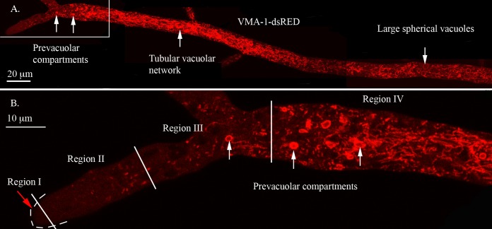
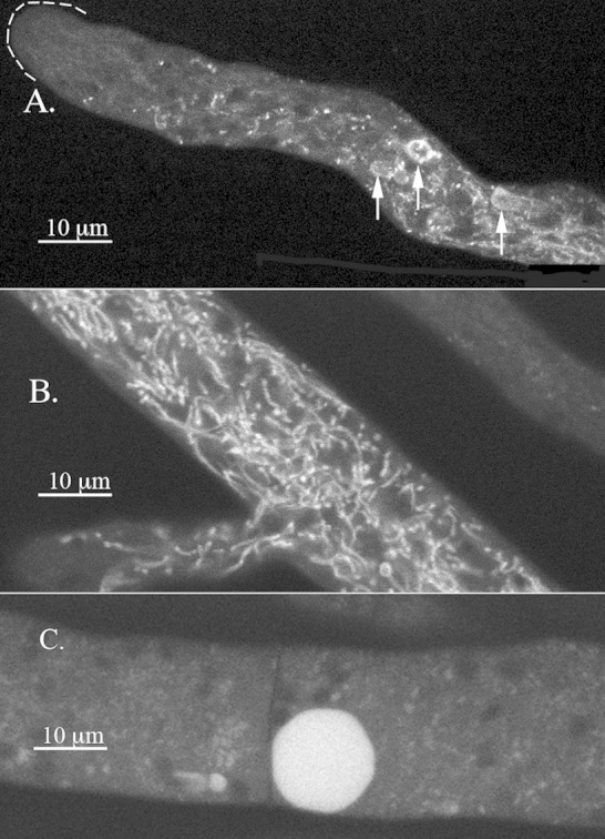
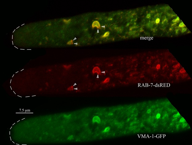
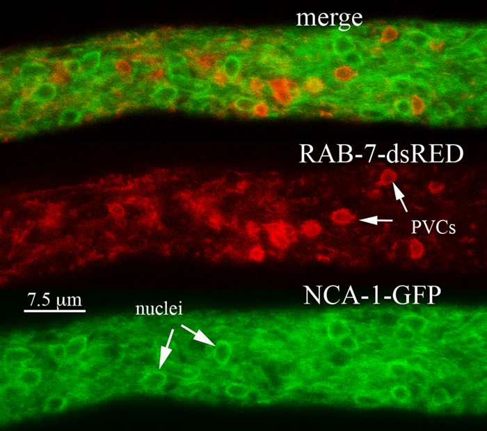
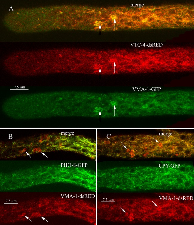
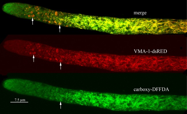
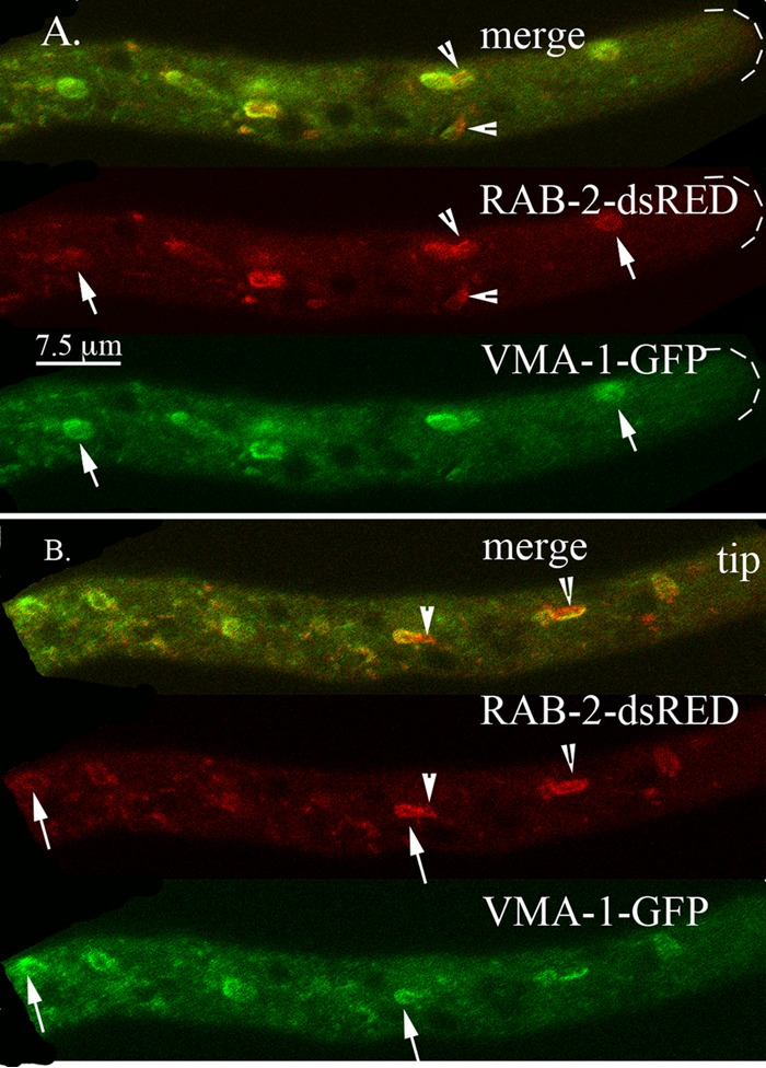
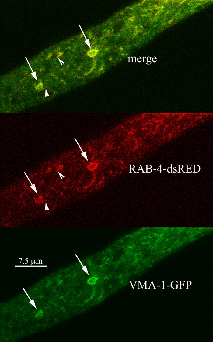
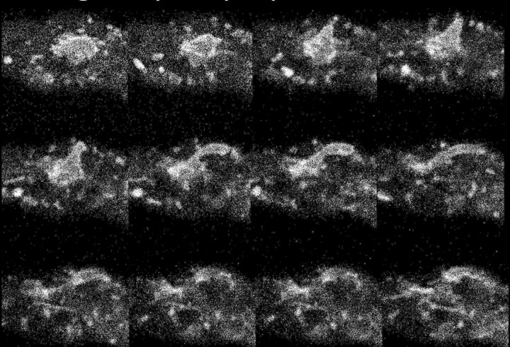
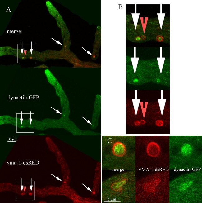
Using confocal microscopy, we observed ring-like organelles, similar in size to nuclei, in the hyphal tip of the filamentous fungus Neurospora crassa. These organelles contained a subset of vacuolar proteins. We hypothesize that they are novel prevacuolar compartments (PVCs). We examined the locations of several vacuolar enzymes and of fluorescent compounds that target the vacuole. Vacuolar membrane proteins, such as the vacuolar ATPase (VMA-1) and the polyphosphate polymerase (VTC-4), were observed in the PVCs. A pigment produced by adenine auxotrophs, used to visualize vacuoles, also accumulated in PVCs. Soluble enzymes of the vacuolar lumen, alkaline phosphatase and carboxypeptidase Y, were not observed in PVCs. The fluorescent molecule Oregon Green 488 carboxylic acid diacetate, succinimidyl ester (carboxy-DFFDA) accumulated in vacuoles and in a subset of PVCs, suggesting maturation of PVCs from the tip to distal regions. Three of the nine Rab GTPases in N. crassa, RAB-2, RAB-4, and RAB-7, localized to the PVCs. RAB-2 and RAB-4, which have similar amino acid sequences, are present in filamentous fungi but not in yeasts, and no function has previously been reported for these Rab GTPases in fungi. PVCs are highly pleomorphic, producing tubular projections that subsequently become detached. Dynein and dynactin formed globular clusters enclosed inside the lumen of PVCs. The size, structure, dynamic behavior, and protein composition of the PVCs appear to be significantly different from those of the well-studied prevacuolar compartment of yeasts.


Figures

FIG 1
FIG 1
Visualization of the vacuolar ATPase in different regions of the hypha. The hypha was transformed with VMA-1–dsRED. (A) Composite image made from four overlapping photos of the hypha. The structures seen in different regions are described in the text. (B) Enlargement of the boxed area of the hyphal tip in panel A. Regions I, II, III, and IV are separated by white lines, with white arrows pointing to examples of PVCs.
FIG 2
FIG 2
Visualization of the pigment in the adenine auxotroph ad-3B strain grown on medium with 0.1 mM adenine. (A) Hyphal tip region with arrows pointing to the PVCs. (B) Region of the hypha ∼200 μm from the tip where the tubular vacuolar network appears. (C) Region of the hypha several millimeters from the tip with a large spherical vacuole.
FIG 3
FIG 3
Localization of Rab-7 GTPase (RAB-7) compared to the localization of the vacuolar ATPase (VMA-1) at the hyphal tip. Some small red puncta, marked by arrowheads, occur at the edges of the PVCs.
FIG 4
FIG 4
Localization of the nuclear envelope and endoplasmic reticulum with NCA-1–GFP compared with the localization of RAB-7–dsRED. The region of the hypha shown is ∼100 μm from the hyphal tip.
FIG 5
FIG 5
(A) Localization of the vacuolar polyphosphate polymerase (VTC-4) compared to the localization of the vacuolar ATPase (VMA-1). (B) Localization of the vacuolar alkaline phosphatase (PHO-8) compared to the localization of the vacuolar ATPase (VMA-1). (C) Localization of carboxypeptidase Y (CPY) compared to the localization of the vacuolar ATPase (VMA-1). In each panel, arrows point to examples of PVCs.
FIG 6
FIG 6
Visualization of carboxy-DFFDA a soluble dye that localizes to vacuoles, compared to the localization of the vacuolar ATPase (VMA-1). The VMA-1–dsRED strain was incubated with carboxy-DFFDA as described in Materials and Methods. Arrows point to examples of PVCs.
FIG 7
FIG 7
Localization of the Rab-2 GTPase (RAB-2) compared to the localization of the vacuolar ATPase (VMA-1). Arrows point to examples of PVCs. Arrowheads point to structures that contain both RAB-2–dsRED and VMA-1–GFP. Panels A and B show examples of different hyphae from the same heterokaryon.
FIG 8
FIG 8
Localization of the Rab-4 GTPase (RAB-4) compared to the localization of the vacuolar ATPase (VMA-1). Some small red puncta, marked by arrowheads, were observed at the edges of the PVCs.
FIG 9
FIG 9
Changes in the shape of PVCs visualized with RAB-7–dsRED. The figure shows successive frames taken at 5.4-s intervals from a movie (see Movie S3 in the supplemental material).
FIG 10
FIG 10
Localization of dynactin compared to the localization of the vacuolar ATPase (VMA-1). (A) Low-magnification image of the heterokaryon. Arrows point to the PVCs. The red arrowhead points to a PVC that does not appear to contain dynactin-GFP. (B) Boxed regions of panel A at a higher magnification. (C) Two PVCs from other hyphae at a higher magnification.

Similar articles

Cited by

References

    1. Bowman EJ, Bowman BJ. 2010. Vacuoles in filamentous fungi, p 179–190. In Borkovich K, Ebbole DJ (ed), Cellular and molecular biology of filamentous fungi. ASM Press, Washington, DC.
    1. Klionsky DJ, Herman PK, Emr SD. 1990. The fungal vacuole: composition, function, and biogenesis. Microbiol Rev 54:266–292. - PMC - PubMed
    1. Richards A, Veses V, Gow NAR. 2010. Vacuole dynamics in fungi. Fungal Biol Rev 24:93–105. doi:10.1016/j.fbr.2010.04.002. - DOI
    1. Veses V, Richards A, Gow NA. 2008. Vacuoles and fungal biology. Curr Opin Microbiol 11:503–510. doi:10.1016/j.mib.2008.09.017. - DOI - PubMed
    1. Li SC, Kane PM. 2009. The yeast lysosome-like vacuole: endpoint and crossroads. Biochim Biophys Acta 1793:650–663. doi:10.1016/j.bbamcr.2008.08.003. - DOI - PMC - PubMed

Publication types

MeSH terms