. 2016 Jan;283(1):88-101.
doi: 10.1111/febs.13553. Epub 2015 Oct 31.

DDX3 functions in antiviral innate immunity through translational control of PACT

Affiliations

DDX3 functions in antiviral innate immunity through translational control of PACT

Ming-Chih Lai et al. FEBS J. 2016 Jan.

Abstract

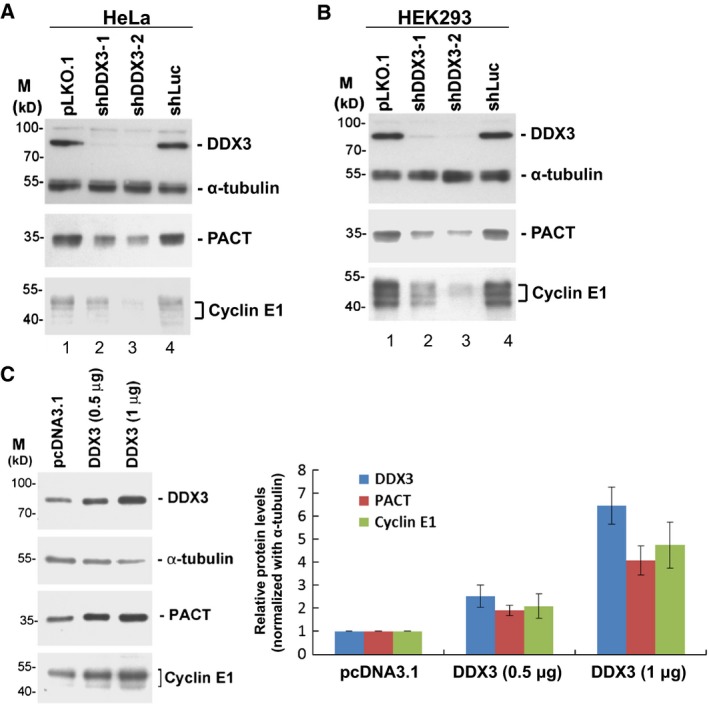
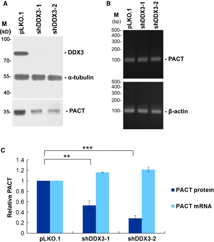
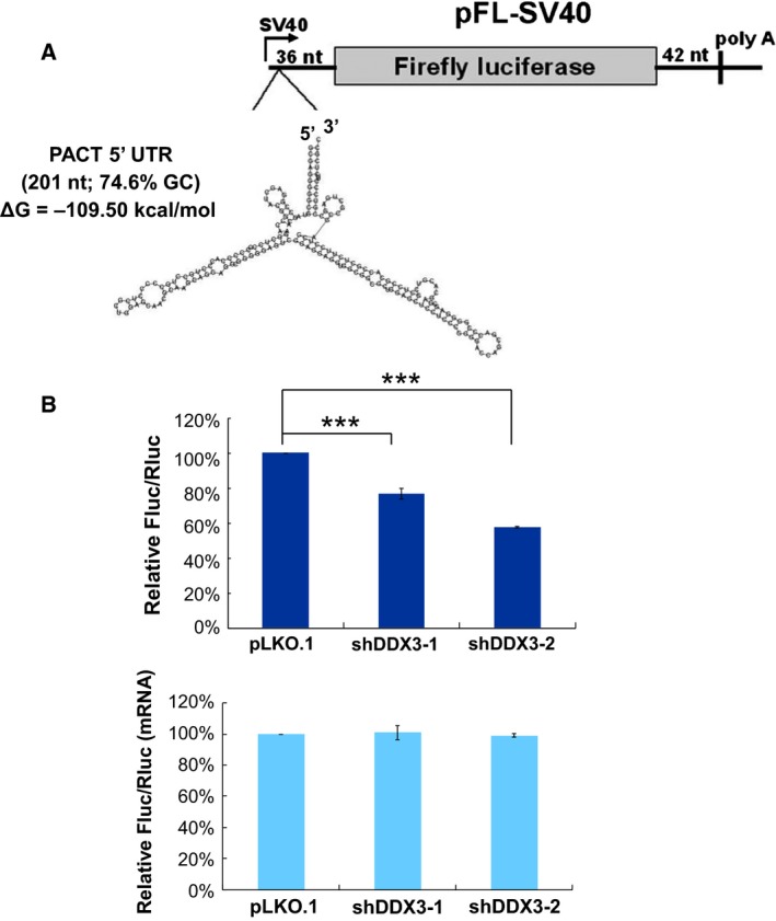
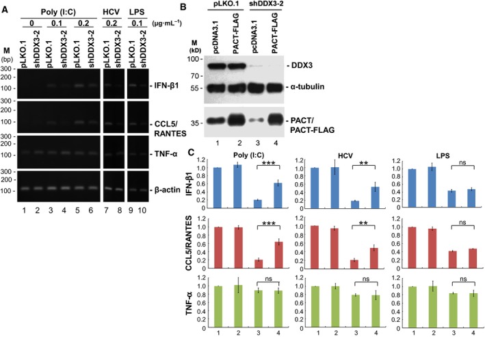
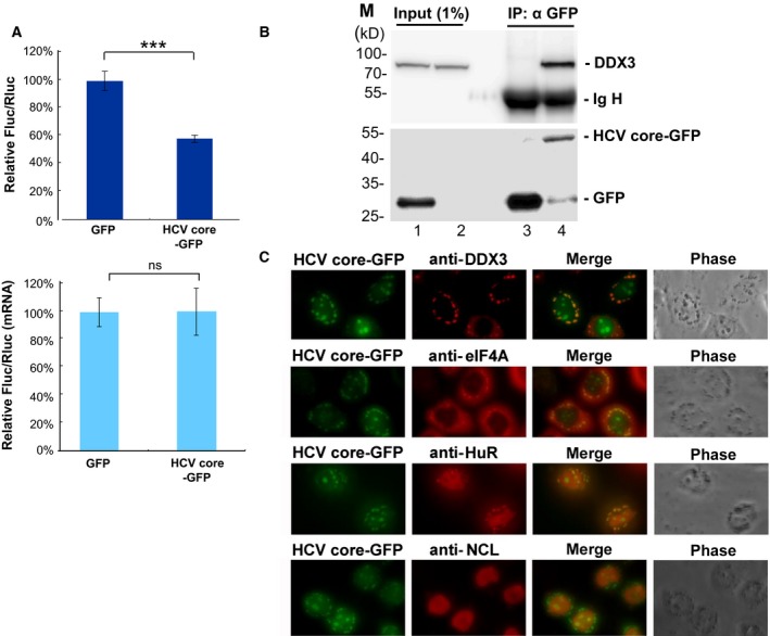
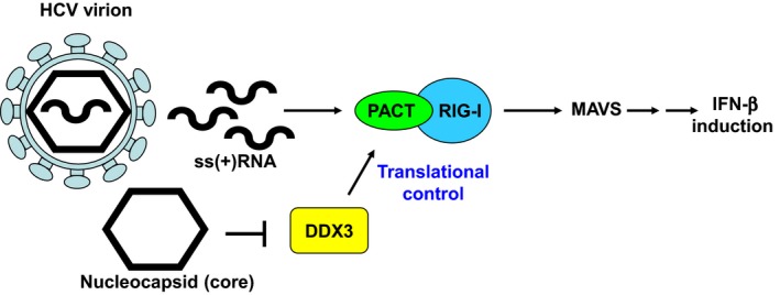
It has emerged that DDX3 plays a role in antiviral innate immunity. However, the exact mechanism by which DDX3 functions in antiviral innate immunity remains to be determined. We found that the expression of the protein activator of the interferon-induced protein kinase (PACT) was regulated by DDX3 in human cells. PACT acts as a cellular activator of retinoic acid-inducible gene-I-like receptors in the sensing of viral RNAs. DDX3 facilitated the translation of PACT mRNA that may contain a structured 5' UTR. Knockdown of DDX3 decreased the viral RNA detection sensitivity of the cells. PACT partially rescued defects of interferon-β1 and chemokine (C-C motif) ligand 5/RANTES (regulated on activation normal T cell expressed and secreted) induction in DDX3-knockdown HEK293 cells. Therefore, DDX3 may participate in antiviral innate immunity, at least in part, by translational control of PACT. Moreover, we show that overexpression of the hepatitis C virus (HCV) core protein inhibited the translation of a reporter mRNA harboring the PACT 5' UTR. The HCV core protein was associated and colocalized with DDX3 in cytoplasmic stress granules, suggesting that the HCV core may abrogate the function of DDX3 by sequestering DDX3 in stress granules. The perturbation of DDX3 by viral proteins delineates a critical role for DDX3 in antiviral host defense.

Keywords: DDX3; PACT; antiviral innate immunity; hepatitis C virus; translational control.


Figures

Figure 1
Figure 1
DDX3 regulates the expression of PACT. (A) HeLa cells were transduced with the empty lentiviral vector (lane 1, pLKO.1) or the pLKO.1 vector expressing the indicated shRNAs (lanes 2–4). After 24 h, puromycin was added to the culture medium for selection. Cells were harvested for analysis at 3 days post‐transduction. Immunoblotting was performed using antibodies against DDX3, α‐tubulin, PACT and cyclin E1. Detection of α‐tubulin was used as a loading control. (B) HEK293 cells were transduced with the lentiviral vectors as described in (A), except that puromycin was omitted. (C) HeLa cells were transfected with the pcDNA3.1 vector or the DDX3‐expressing pcDNA3.1 vector (0.5 and 1 μg). After 48 h, cells were harvested for immunoblotting analysis using antibodies against DDX3, α‐tubulin, PACT and cyclin E1. The bar graph shows the relative expression levels of DDX3, PACT and cyclin E1 normalized to α‐tubulin as the mean ± SEM of at least three independent experiments.
Figure 2
Figure 2
Knockdown of DDX3 inhibits PACT expression at the protein level. (A) HeLa cells were transduced with the empty lentiviral vector (pLKO.1) or the pLKO.1 vector expressing DDX3 shRNAs (shDDX3‐1 and shDDX3‐2). After 24 h, puromycin was added to the culture for selection. Cells were harvested for analysis at 3 days post‐transduction. Immunoblotting was performed using antibodies against DDX3, α‐tubulin and PACT. Detection of α‐tubulin was used as a loading control. (B) RNA extracted from mock‐treated (pLKO.1) or DDX3‐knockdown (shDDX3‐1 and shDDX3‐2) HeLa cells was analyzed by conventional RTPCR using specific primers for PACT and β‐actin mRNAs. PCR products were resolved by agarose gel electrophoresis. (C) The protein level of PACT relative to α‐tubulin in mock‐treated (pLKO.1) and DDX3‐knockdown (shDDX3‐1 and shDDX3‐2) HeLa cells were quantitatively analyzed. The levels of PACT and β‐actin mRNAs were detected by quantitative real‐time RTPCR. The bar graph shows the relative expression levels of PACT protein and mRNA as the mean ± SEM of at least three independent experiments (**P < 0.01, ***P < 0.001).
Figure 3
Figure 3
Knockdown of DDX3 inhibits translation initiation of PACT mRNA. (A) HeLa cells were transduced with the empty lentiviral vector (pLKO.1) or the pLKO.1 vector expressing DDX3 shRNAs (shDDX3‐1 and shDDX3‐2). Cells were harvested for analysis at 3 days post‐transduction. Cytoplasmic extracts were loaded on a linear 15–40% sucrose gradient ultracentrifugation. After centrifugation, the polysome profile was plotted by A 254 values (top). Total RNA was extracted from each fraction for analysis. The purified RNA was resolved on a 1% formaldehyde/agarose gel, and rRNAs were visualized by ethidium bromide staining (bottom). The levels of mRNA were analyzed by quantitative real‐time RTPCR using specific primers for PACT, cyclin E1 and β‐actin mRNAs (middle). (B) Translational efficiency of β‐actin, PACT and cyclin E1 mRNAs in mock‐treated (pLKO.1) and DDX3‐knockdown (shDDX3‐1 and shDDX3‐2) HeLa cells was calculated and shown as a percentage. The bar graph shows the changes of translational efficiency as the mean ± SEM of at least three independent experiments (ns, not significant; ***P < 0.001, *P < 0.05).
Figure 4
Figure 4
DDX3 facilitates the translation of reporter mRNAs containing the PACT 5′ UTR. (A) Schematic representation of the firefly luciferase (Fluc) reporter (pFL‐SV40) with the human PACT 5′ UTR (201 nucleotides), for which the predicted secondary structure is shown. (B) HeLa cells were transduced with the empty lentiviral vector (pLKO.1) or the pLKO.1 vector expressing DDX3 shRNAs (shDDX3‐1 and shDDX3‐2). After 48 h, HeLa cells were co‐transfected with the pFL‐SV40 reporter containing the PACT 5′ UTR and the control pRL‐SV40 vector encoding the Renilla luciferase (Rluc). Cells were lysed for analysis at 24 h post‐transfection. For each transfectant, the Fluc activity was normalized to that of the Rluc control. The bar graph shows the relative Fluc/Rluc activities in DDX3‐knockdown cells compared to mock‐treated cells (top). Fluc and Rluc mRNAs were analyzed by quantitative real‐time RTPCR (bottom). Data are shown as the mean ± SEM from at least three independent experiments (***P < 0.001).
Figure 5
Figure 5
PACT partially rescued the antiviral innate immune defects caused by DDX3 knockdown. HEK293 cells were transduced with the empty lentiviral vector (pLKO.1) or the pLKO.1 vector expressing DDX3 shRNA (shDDX3‐2) for subsequent functional assessment. (A) At 3 days post‐transduction, HEK293 cells were transfected with poly(I:C) or the HCV PAMP RNA or treated with bacterial LPS for 4 h at different concentrations as indicated (μg·mL−1). Total RNA extracted from HEK293 cells was analyzed by conventional RTPCR using specific primers for IFN‐β1, CCL5/RANTES, TNF‐α, and β‐actin mRNAs. PCR products were resolved by 1.5% agarose gel electrophoresis. (B) At 48 h post‐transduction, cells were further transfected with the pcDNA3.1 vector or the FLAG‐tagged PACT‐expressing pcDNA3.1 vector (PACTFLAG) to assess whether PACT can be rescued in DDX3‐knockdown cells. Cell lysates were subjected to immunoblotting analysis using antibodies against DDX3, α‐tubulin and PACT. Detection of α‐tubulin was used as a loading control. (C) Under a similar experimental setting to that described in (B), HEK293 transfectants at 3 days post‐transduction were further transfected with 0.1 μg·mL−1 of poly(I:C) or 0.2 μg·mL−1 of the HCV PAMP RNA, or treated with 0.1 μg·mL−1 of bacterial LPS for 4 h to induce antiviral innate immune response. Total RNA extracted from HEK293 cells was analyzed by quantitative real‐time RTPCR using specific primers for IFN‐β1, CCL5/RANTES, TNF‐α and β‐actin mRNAs. The bar graph shows the relative mRNA levels of IFN‐β1, CCL5/RANTES and TNF‐α normalized to β‐actin as the mean ± SEM from at least three independent experiments (***P < 0.001, **P < 0.01; ns, not significant).
Figure 6
Figure 6
HCV core protein inhibits PACT translation by direct binding and redistribution of DDX3 into cytoplasmic SGs. (A) HeLa cells were co‐transfected with the pFL‐SV40 reporter containing the PACT 5′ UTR and the control pRL‐SV40 vector in combination with the pEGFP‐N1 vector encoding GFP or HCV core‐GFP. Cells were lysed for analysis at 48 h post‐transfection. For each transfectant, the Fluc activity was normalized to that of the Rluc control. The bar graph shows the relative Fluc/Rluc activities in HCV core‐overexpressing (HCV core‐GFP) cells compared to control (GFP) cells (top). Fluc and Rluc mRNAs were analyzed by quantitative real‐time RTPCR (bottom). Data are shown as the mean ± SEM from at least three independent experiments (***P < 0.001; ns, not significant). (B) HEK293 cells were transfected with the pEGFP‐N1 vector encoding GFP or HCV core‐GFP. Cells were lysed for analysis at 48 h post‐transfection. Immunoprecipitation was performed using anti‐GFP antibody coupled to protein A sepharose beads. Immunoprecipitates were treated with 1 mg·mL−1 RNase A at 37 °C for 30 min. Bound proteins were eluted and subjected to immunoblotting with anti‐DDX3 antibody (top) or anti‐GFP antibody (bottom). Ig H represents the immunoglobulin heavy chain. (C) HeLa cells were transfected with the pEGFP‐N1 vector encoding HCV core‐GFP for 24 h. Immunofluorescent staining of HeLa cells was carried out using antibodies against DDX3, eIF4A, HuR and nucleolin (NCL). Co‐localization of HCV core‐GFP (green) and DDX3, eIF4A or HuR (red) in cytoplasmic SGs was observed under a fluorescence microscope.
Figure 7
Figure 7
A model of DDX3‐mediated translational control of PACT in antiviral innate immune response to HCV infection. The HCV genomic RNA (ssRNA) contains specific PAMPs, which can be detected by RIG‐I to trigger antiviral innate immunity (IFN‐β induction). The dsRNA binding protein PACT is a binding partner of RIG‐I and functions as an activator of RIG‐I in the sensing of HCV RNAs. Interestingly, the HCV core protein abrogates the RIG‐I‐mediated antiviral innate immune response by direct binding of DDX3. Inactivation of DDX3 down‐regulates the expression of PACT and thus attenuates the viral RNA detection sensitivity of the cells.

Similar articles

Cited by

References

    1. Cordin O, Banroques J, Tanner NK & Linder P (2006) The DEAD‐box protein family of RNA helicases. Gene 367, 17–37. - PubMed
    1. Rocak S & Linder P (2004) DEAD‐box proteins: the driving forces behind RNA metabolism. Nat Rev Mol Cell Biol 5, 232–241. - PubMed
    1. Botlagunta M, Vesuna F, Mironchik Y, Raman A, Lisok A, Winnard P Jr, Mukadam S, Van Diest P, Chen JH, Farabaugh P et al (2008) Oncogenic role of DDX3 in breast cancer biogenesis. Oncogene 27, 3912–3922. - PMC - PubMed
    1. Chao CH, Chen CM, Cheng PL, Shih JW, Tsou AP & Lee YH (2006) DDX3, a DEAD box RNA helicase with tumor growth‐suppressive property and transcriptional regulation activity of the p21waf1/cip1 promoter, is a candidate tumor suppressor. Cancer Res 66, 6579–6588. - PubMed
    1. Jamieson DJ, Rahe B, Pringle J & Beggs JD (1991) A suppressor of a yeast splicing mutation (prp8‐1) encodes a putative ATP‐dependent RNA helicase. Nature 349, 715–717. - PubMed

Publication types