. 2015 Dec:6:516-523.
doi: 10.1016/j.redox.2015.06.016. Epub 2015 Jun 27.

Autophagy in neonatal hypoxia ischemic brain is associated with oxidative stress

Affiliations

Autophagy in neonatal hypoxia ischemic brain is associated with oxidative stress

Qing Lu et al. Redox Biol. 2015 Dec.

Abstract

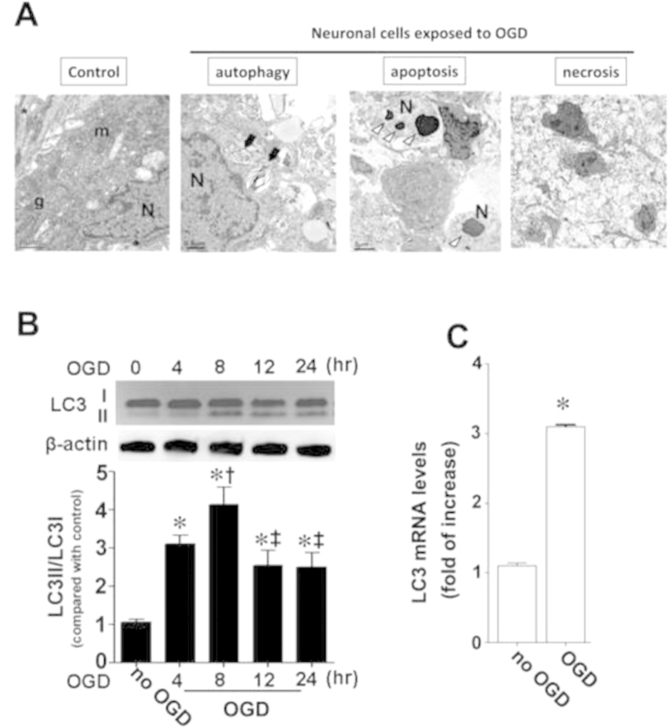
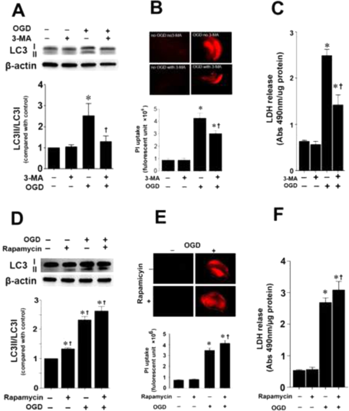
Autophagy is activated when the neonatal brain exposed to hypoxia ischemia (HI), but the mechanisms underlying its activation and its role in the neuronal cell death associated with HI is unclear. We have previously shown that reactive oxygen species (ROS) derived from nicotinamide adenine dinucleotide phosphate (NADPH) oxidase play an important role in HI-mediated neuronal cell death. Thus, the aim of this study was to determine if ROS is involved in the activation of autophagy in HI-mediated neonatal brain injury and to determine if this is a protective or deleterious pathway. Initial electron microscopy data demonstrated that autophagosome formation is elevated in P7 hippocampal slice cultures exposed to oxygen-glucose deprivation (OGD). This corresponded with increased levels of LC3II mRNA and protein. The autophagy inhibitor, 3-methyladenine (3-MA) effectively reduced LC3II levels and autophagosome formation in hippocampal slice cultures exposed to OGD. Neuronal cell death was significantly attenuated. Finally, we found that the pharmacologic inhibition of NADPH oxidase using apocynin or gp91ds-tat decreased autophagy in hippocampal slice cultures and the rat brain respectively. Thus, our results suggest that an activation of autophagy contributes to neonatal HI brain injury this is oxidative stress dependent.

Keywords: Autophagy; Hypoxia–ischemia; NADPH oxidase; Neonatal brain; Neuronal cell death.


Figures

None
Graphical abstract
Fig. 1
Fig. 1
Autophagy is induced in rat hippocampal slice cultures exposed to oxygen glucose deprivation. Transmission electron microscopy identifies autophagosome formation (black arrow) in the neuronal cell as well as markers of apoptosis (white triangle) and necrosis, in hippocampal slice cultures 8 h after OGD. m, Mitochondria; N, nuclei; g, golgi apparatus. (A) Rat hippocampal slice cultures were harvested 4-, 8-, 12- and 24 h after OGD to determine LC3II protein (B) and mRNA levels (C). OGD increases both LC3II protein (B) and mRNA (C). Data are presented as mean±S.E from 4 independent experiments using 24 pooled slices per experiment. *P<0.05 vs. control, P<0.05 vs. 4 h OGD, P<0.05 vs. 8 h OGD.
Fig. 2
Fig. 2
Modulation of autophagy alters neuronal cell death in rat hippocampal slice cultures exposed to oxygen glucose deprivation. Rat hippocampal slice cultures were exposed to OGD in the presence of the autophagy inhibitor 3-MA (5 mM, 2 h prior to OGD), or the autophagy activator, rapamycin (200 nM), both given 2 h prior to OGD. Slices were harvested 8 h after OGD and subjected to Western blot analysis to determine effects on LC3II (A and D), whole slice PI uptake (B and E) and LDH release levels (C and F). 3-MA attenuated LC3II protein levels (A), PI uptake (B) and LDH release (C). Rapamycin induced the opposite effects (D–F). Data are presented as mean±S.E from 4 independent experiments using 24 pooled slices per experiment. *P<0.05 vs. no OGD, P<0.05 vs. OGD only.
Fig. 3
Fig. 3
NADPH oxidase inhibition reduces autophagy in OGD exposed hippocampal slice cultures. Rat hippocampal slice cultures were exposed to OGD in the presence of the NADPH oxidase inhibitor, apocynin (100 µM), 2 h prior to OGD, then harvested at 8 h after OGD. Western blot analysis was used to determine the effect on the LC3II levels (A). Values are presented as mean±S.E from 4 independent experiments using 24 pooled slices per experiment. *P<0.05 vs. no OGD, P<0.05 vs. no treatment under OGD. Apocynin also reduced autophagosomes formation as determined using both fluorescent- (B) and transmission electron-microscopy (C). Values are presented as mean±S.E from 4 independent experiments. *P<0.05 vs. no OGD no treatment, P<0.05 vs. no OGD with treatment.
Fig. 4
Fig. 4
NADPH oxidase inhibition attenuates autophagy in the neonatal rat brain exposed to hypoxia–ischemia. P7 neonatal rats were pre-treated with gp91ds-tat, the scrambled control peptide, or vehicle then exposed to HI. Two hours after HI, LC3II protein levels and tissue autophagosome formation were analyzed. There is a significant increase in the LC3II/LC3I protein ratio in the right hemisphere of the neonatal brain and this is attenuated by gp91ds-tat (A). The increase in autophagosome formation in the right hemisphere of the neonatal brain, as determined using fluorescent- (B and C) and transmission electron-microscopy (D), is also attenuated by gp91ds-tat. Values are presented as mean±S.E from 6 animals per group. *P<0.05 vs. left hemisphere, P<0.05 vs. HI+vehicle, P<0.05 vs. HI+ScrTAT.

References

    1. Ferriero D.M. Neonatal brain injury. N. Engl. J. Med. 2004;351:1985–1995. - PubMed
    1. Odding E., Roebroeck M.E., Stam H.J. The epidemiology of cerebral palsy: incidence, impairments and risk factors. Disabil. Rehabil. 2006;28:183–191. - PubMed
    1. Roth S.C., Baudin J., Cady E., Johal K., Townsend J.P., Wyatt J.S., Reynolds E.O., Stewart A.L. Relation of deranged neonatal cerebral oxidative metabolism with neurodevelopmental outcome and head circumference at 4 years. Dev. Med. Child Neurol. 1997;39:718–725. - PubMed
    1. Blumberg R.M., Cady E.B., Wigglesworth J.S., McKenzie J.E., Edwards A.D. Relation between delayed impairment of cerebral energy metabolism and infarction following transient focal hypoxia–ischaemia in the developing brain. Exp. Brain Res. 1997;113:130–137. - PubMed
    1. Lu Q., Rau T.F., Harris V., Johnson M., Poulsen D.J., Black S.M. Increased p38 mitogen-activated protein kinase signaling is involved in the oxidative stress associated with oxygen and glucose deprivation in neonatal hippocampal slice cultures. Eur. J. Neurosci. 2011;34:1093–1101. - PMC - PubMed

Publication types

LinkOut - more resources