Review
. 2016 Mar;468(3):259-75.
doi: 10.1007/s00428-015-1858-9. Epub 2015 Oct 10.

The many faces of small B cell lymphomas with plasmacytic differentiation and the contribution of MYD88 testing

Affiliations
Review

The many faces of small B cell lymphomas with plasmacytic differentiation and the contribution of MYD88 testing

Steven H Swerdlow et al. Virchows Arch. 2016 Mar.

Abstract

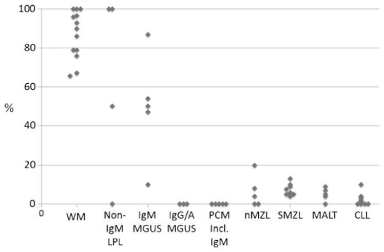
Plasmacytic differentiation may occur in almost all small B cell lymphomas (SBLs), although it varies from being uniformly present (as in lymphoplasmacytic lymphoma (LPL)) to very uncommon (as in mantle cell lymphomas (MCLs)). The discovery of MYD88 L265P mutations in the vast majority of LPLs has had a major impact on the study of these lymphomas. Review of the cases contributed to the 2014 European Association for Haematopathology/Society for Hematopathology slide workshop illustrated how mutational testing has helped refine the diagnostic criteria for LPL, emphasizing the importance of identifying a clonal monotonous lymphoplasmacytic population and highlighting how LPL can still be diagnosed with extensive nodal architectural effacement, very subtle plasmacytic differentiation, follicular colonization, or uncommon phenotypes such as CD5 or CD10 expression. MYD88 L265P mutations were found in 11/11 LPL cases versus only 2 of 28 other SBLs included in its differential diagnosis. Mutational testing also helped to exclude other cases that would have been considered LPL in the past. The workshop also highlighted how plasmacytic differentiation can occur in chronic lymphocytic leukemia/small lymphocytic lymphoma, follicular lymphoma, SOX11 negative MCL, and particularly in marginal zone lymphomas, all of which can cause diagnostic confusion with LPL. The cases also highlighted the difficulty in distinguishing lymphomas with marked plasmacytic differentiation from plasma cell neoplasms. Some SBLs with plasmacytic differentiation can be associated with amyloid, other immunoglobulin deposition, or crystal-storing histiocytosis, which may obscure the underlying neoplasm. Finally, although generally indolent, LPL may transform, with the workshop cases suggesting a role for TP53 abnormalities.

Keywords: Chronic lymphocytic leukemia; Follicular lymphoma; Lymphoplasmacytic lymphoma; MYD88; Mantle cell lymphoma; Marginal zone lymphoma; Plasmacytic differentiation.


Figures

Fig. 1
Fig. 1
CD10+ lymphoplasmacytic lymphoma with MYD88 L265P mutation and typical cytogenetic findings. a Note the monotonous cell population between the widely patent sinuses, typical for LPL. b There are many small lymphoid cells admixed with obvious plasmacytoid and plasma cells. The infiltrate is c CD20+ and d CD10+. e Classical cytogenetics demonstrated 47,XX,+4,i(6)(p10) in 6 of 19 cells. The latter abnormality is equivalent to deletion 6q (case 267, courtesy of K. Gill)
Fig. 2
Fig. 2
Frequency of MYD88 L265P mutations in B cell neoplasms. Compilation of numerous studies reported in the literature demonstrates that many but not all LPL have MYD88 L265P mutations even if they are not IgM+. Some cases of IgM MGUS are mutated but not IGG/A MGUS nor myeloma even if IgM+. A small proportion of other small B cell lymphomas are mutated (see text). In addition, up to about one third of non-GC/ABC-type DLBCL have MYD88 L265P mutations, with a higher proportion of leg-type DLBCL, CNS, and testicular DLBCL (not illustrated in figure). References available upon request
Fig. 3
Fig. 3
CD5+ lymphoplasmacytic lymphoma with MYD88 L265P mutation and limited morphologic plasmacytic features. a Note the monotonous cell population between the widely patent sinuses, typical for LPL. b There are numerous small lymphoid cells and focal hemosiderin. c Tryptase stain highlights prominent mast cells. The B cell population is d CD3−, e CD5+, and f LEF1− (positive T cells are present). g An IgM stain highlights the mostly perisinus plasmacytic component (case 190, courtesy of X. Zhao et al.)
Fig. 4
Fig. 4
a–d Lymphoplasmacytic lymphoma with MYD88 L265P mutation, marked follicular colonization, and extensive architectural effacement. a There is architectural effacement with some hyalinized small vessels and other sclerosis. b The CD21 immunohistochemical stain highlights the striking follicular colonization. c Small lymphocytes with admixed plasmacytoid and plasmacytic cells as well as hemosiderin are present. d The kappa stain showed varying degrees of cytoplasmic positivity and highlighted Dutcher bodies (arrows). A lambda stain showed only non-specific staining (not shown) (case 80, courtesy of L. Venkatraman et al.). e Favor mantle cell lymphoma (cyclin D1+ but without demonstrable CCND1 rearrangement) with plasmacytic differentiation. Note how easily this lymphoma could be confused with a lymphoplasmacytic lymphoma, with its lymphoplasmacytic proliferation and a Dutcher body (arrow). The cyclin D1 staining was considered incompatible with the diagnosis of lymphoplasmacytic lymphoma or marginal zone lymphoma and the wild-type MYD88 also very consistent with the panel’s diagnosis (case 46, courtesy of M. R. Ashton-Key)
Fig. 5
Fig. 5
Accelerated CLL with plasmacytic differentiation. a Note the prominent proliferation center. Immunohistochemical stains demonstrated b minimal staining for kappa but c numerous cells with cytoplasmic lambda staining and d extensive positivity for BLIMP1 (case 89, courtesy of D. Martinez et al.)
Fig. 6
Fig. 6
Follicular lymphoma with plasmacytic differentiation and BCL2 and BCL6 rearrangements. a Architectural effacement by a somewhat ill-defined follicular proliferation is present. b The neoplastic germinal center includes both Dutcher bodies (short arrow) and Mott cells (longer arrow). c A CD21 stain confirms the follicular architecture. d The CD138 stain highlights the prominent intrafollicular and interfollicular plasmacytic population. There is cytoplasmic e IgM- and f kappa-restricted positivity. Both IgM and kappa stains highlighted the presence of Dutcher bodies (arrows) (case 150, courtesy of M. Kinney)
Fig. 7
Fig. 7
Mantle cell lymphoma with plasmacytic differentiation and MYD88 mutation. a Mantle cell lymphoma with a residual naked germinal center. b The cells were small with a round nucleus, and some had Dutcher bodies (arrow). c The neoplastic cells were SOX11-negative but d strongly positive for cyclin D1. e Cells with Dutcher bodies were also cyclin D1-positive (arrow) (case 89, courtesy of I. Ribera-Cortada et al.)
Fig. 8
Fig. 8
Nodal marginal zone lymphomas with plasmacytic differentiation. a Note the typical marginal zone growth pattern in this NMZL with wild-type MYD88 b with numerous CD20 positive B cells c that outside of the germinal center express CD5. d The CD3 highlights the benign T cells. e The IgM immunostain marks both the neoplastic B cells and a small number of neoplastic plasma cells (a–e, case 302, courtesy of S. Isiksoy). (f) This NMZL with a MYD88 L265P mutation also had a typical marginal zone growth pattern. g Note the CD20 positive B cells in the follicle and at the periphery of the image (red), the surrounding CD3 positive T cells (brown), and the presence of cells marking with neither. h A CD138 stain highlights the prominent plasma cells outside the nodules. i There was also focally marked p53 expression (f–i, case 288, courtesy of M. Klimkowska)
Fig. 9
Fig. 9
MALT lymphomas with plasmacytic differentiation. a This MALT lymphoma of the thyroid from a 52-year-old man with a history of Hashimoto’s thyroiditis had b very limited CD20 but c marked CD79a expression. d The plasmacytic cells marked with neither kappa or lambda in this double in situ hybridization stain but e showed extensive IgG positivity (a–e, case 270, courtesy of E. M. Maggio). f This MALT lymphoma that was associated with chronic sclerosing sialadenitis had g, h kappa monotypic plasma cells with i about 36 % positive for IgG4 (f–i, case 264, courtesy of J. M. Jaso). j This dural MALT lymphoma included numerous plasma cells that k were kappa monotypic (inset lambda) based on in situ hybridization stains and were l IgG and m IgG4 positive (case 255, courtesy of T. Gindin)
Fig. 10
Fig. 10
Splenic marginal zone lymphoma with plasmacytic differentiation and wild-type MYD88. a Note the typical biphasic pattern but with b focally numerous plasma cells that were c IgM positive (case 178, courtesy of M. A. Piris)
Fig. 11
Fig. 11
Histologic, immunophenotypic, and molecular features of transformed LPL. a Axillary lymph node showing architectural distortion by small lymphocytes, plasmacytoid lymphocytes, and plasma cells. b Focally, there are clusters of large atypical cells with open chromatin and conspicuous nucleoli. Mitotic figures are prominent. c There are more abundant p53-positive cells in the large-cell component (right half). d Targeted massively parallel sequencing reveals the characteristic point mutation in MYD88 (L265P) with an allele frequency of 28 % and e a deletion of three nucleotides in TP53 (V218del) with an allele frequency of 49 % (case 144, courtesy of C. Soderquist)

References

    1. Andriko JA, Swerdlow SH, Aguilera NI, Abbondanzo SL. Is lymphoplasmacytic lymphoma/immunocytoma a distinct entity? A clinicopathologic study of 20 cases. Am J Surg Pathol. 2001;25:742–751. - PubMed
    1. Bob R, Falini B, Marafioti T, Paterson JC, Pileri S, Stein H. Nodal reactive and neoplastic proliferation of monocytoid and marginal zone B cells: an immunoarchitectural and molecular study highlighting the relevance of IRTA1 and T-bet as positive markers. Histopathology. 2013;63:482–498. doi: 10.1111/his.12160. - DOI - PubMed
    1. Brenner I, Roth S, Puppe B, Wobser M, Rosenwald A, Geissinger E. Primary cutaneous marginal zone lymphomas with plasmacytic differentiation show frequent IgG4 expression. Mod Pathol. 2013;26:1568–1576. doi: 10.1038/modpathol.2013.106. - DOI - PubMed
    1. Campo E, Miquel R, Krenacs L, Sorbara L, Raffeld M, Jaffe ES. Primary nodal marginal zone lymphomas of splenic and MALT type. Am J Surg Pathol. 1999;23:59–68. - PubMed
    1. Cheuk W, Yuen HK, Chan AC, Shih LY, Kuo TT, Ma MW, Lo YF, Chan WK, Chan JK. Ocular adnexal lymphoma associated with IgG4+ chronic sclerosing dacryoadenitis: a previously undescribed complication of IgG4-related sclerosing disease. Am J Surg Pathol. 2008;32:1159–1167. doi: 10.1097/PAS.0b013e31816148ad. - DOI - PubMed

MeSH terms

Substances