Review
. 2015 Dec;19(12):783-796.
doi: 10.1016/j.tics.2015.09.002. Epub 2015 Oct 7.

Who is That? Brain Networks and Mechanisms for Identifying Individuals

Affiliations
Review

Who is That? Brain Networks and Mechanisms for Identifying Individuals

Catherine Perrodin et al. Trends Cogn Sci. 2015 Dec.

Abstract

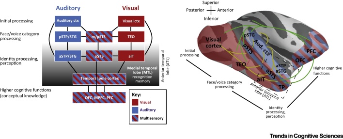
Social animals can identify conspecifics by many forms of sensory input. However, whether the neuronal computations that support this ability to identify individuals rely on modality-independent convergence or involve ongoing synergistic interactions along the multiple sensory streams remains controversial. Direct neuronal measurements at relevant brain sites could address such questions, but this requires better bridging the work in humans and animal models. Here, we overview recent studies in nonhuman primates on voice and face identity-sensitive pathways and evaluate the correspondences to relevant findings in humans. This synthesis provides insights into converging sensory streams in the primate anterior temporal lobe (ATL) for identity processing. Furthermore, we advance a model and suggest how alternative neuronal mechanisms could be tested.

Keywords: face; human; identity; multisensory; primate; temporal lobe; voice.


Figures

Figure 1
Figure 1
Temporal Lobe Voice Areas in Humans, Monkeys, and Dogs. (A) Voice category-sensitive sites (voice versus nonvoice sounds; blue) in the human temporal lobe or those that are voice-identity sensitive (within category; red). The identified sites are projected onto the surface using pySurfer softwarei and correspond to the identified peak of activity clusters reported in , , , , , , . This representation focuses only on the temporal lobe and the right hemisphere, although, as the original reports show, the left hemisphere also has temporal voice-sensitive regions. For a recent probabilistic map of human voice-category sensitive regions, see . (B) Summary of voice-category and voice-identity sensitive sites in the macaque temporal lobe, obtained from peak activity clusters reported in . Also shown are vocalization-sensitive peak responsive sites (purple) reported in other macaque neuroimaging studies , , . (C) Voice-category sensitive areas in the brains of domesticated dogs , showing a cluster in the anterior temporal lobe. Abbreviations: a, anterior; c, caudal (posterior); ESS, ectosylvian sulcus; m, middle; p, posterior; PAC, primary auditory cortex; r, rostral (anterior); rESG, rostral ectosylvian gyrus; SF, Sylvian fissure; SG, Sylvian gyrus; SSS, suprasylvian sulcus; STG, superior temporal gyrus; STP, supratemporal plane; STS, superior temporal sulcus; TP, temporal pole. Images provided by A. Andics (C).
Figure 2
Figure 2
Voice- and Face-Sensitive Neuronal Responses in Monkeys. (A) Targeting approach for recording from the anterior voice identity-sensitive functional magnetic resonance imaging (fMRI) cluster (red). Multisensory cortex in the upper bank of the superior temporal sulcus (STS) is illustrated in yellow. The fundus and the lower bank of the STS can contain face-sensitive clusters (blue, see main text). (B) Voice-sensitive neurons show a categorical response to monkey vocalizations produced by many different callers (MVocs) that is twofold greater than responses to vocalizations from other animals (AVocs) or nonvoice natural sounds (NSounds) . (C) Units sensitive to voice (caller) identity are often found within the pool of voice category-preferring units. Such units show comparable responses to two different vocalizations (here the response to ‘coo’ and ‘grunt’ calls is averaged) but differential responses to individual callers (caller M1 versus M2) . (D) Voice-sensitive neurons respond selectively to a small subset of the stimuli within the conspecific voices. (E) Voice-sensitive cells appear to be more stimulus selective (i.e., respond well to smaller percentages of the presented voices, [63]) compared with face cells, which tend to respond to approximately 55% of the faces within the face stimuli , , . Modified, with permission, from (A,C).
Figure 3
Figure 3
Neuronal Multisensory Influences and Effective Functional Connectivity in the Monkey Brain. (A) Example of a nonlinear (subadditive) multisensory unit in voice-sensitive cortex: firing rates in response to combined audiovisual stimulation (AV, voice and face) significantly differ from the sum of the responses to the unimodal stimuli (A, auditory; V, visual). (B) Neuronal multisensory influences are prominent in voice-sensitive cortex (anterior supratemporal plane; aSTP) but are qualitatively different from those in the anterior superior temporal sulcus (aSTS). For example, aSTS neurons more often display bimodal responses . (C) A study of effective functional connectivity using combined microstimulation and functional magnetic resonance imaging (fMRI) shows that stimulating voice-sensitive cortex (blue cross) tends to elicit fMRI activity in anterior temporal lobe (ATL) regions . (D) By contrast, stimulating the aSTS also elicits fMRI activity in frontal cortex, in particular the orbitofrontal cortex (OFC). Abbreviations: A, anterior; I, inferior; P, posterior; S, superior. Modified, with permission, from (A).
Figure 4
Figure 4
Anterior Temporal Lobe (ATL) Neuronal Recordings in Humans. Intracranial human recordings from several areas in the temporal lobe during an auditory and visual identity naming task. (A) Regions of the ATL are responsive to both a picture and the voice of an individual . By contrast, a visual area in the posterior fusiform gyrus (pFG) responds mainly to the picture, and auditory cortex on the superior temporal gyrus (STG) to the sound. Note that verbal naming followed the period of recording in response to the faces and/or voices as stimuli. (B) Some contacts in the two patients (L206, L242, and L258) show unimodal (picture or voice) responses in the ATL, particularly in the beta band. Other contacts show responses to both. Modified, with permission, from .
Figure 5
Figure 5
Key Figure: Primate Model for Identity-Processing and Multisensory Convergence The model focuses on the auditory pathway involved in extracting voice-identity content in communication signals and the analogous visual pathway. The principles would apply to other sensory input streams, although the regions involved may differ. The key features of the model are the initial sensory and category-sensitive processing stages [middle and posterior superior temporal sulcus (m/pSTS); visual area TEO and auditory regions in posterior supratemporal plane (STP)/superior temporal gyrus (STG)]. Multisensory influences are present throughout the visual and auditory pathway, but are thought to be qualitatively different in the STS, in relation to, for example, anterior (a)STP regions, where the auditory modality is dominant , . Identity-related processes would primarily involve anterior temporal lobe (ATL) regions [anterior STP/STG; anterior (a)STS; and anterior inferior temporal cortex (aIT)]. Not illustrated are interactions with medial temporal lobe (MTL) structures, such as the entorhinal cortex and hippocampus, that could support the recognition of familiar individuals. The model is illustrated to the right on a rendered macaque brain to reveal some of the bidirectional pathways of inter-regional connectivity (yellow), as well as some of the feedback projections to auditory and visual processing streams (green). Several multisensory convergence sites are evident, which for identity-related processes in the ATL appear to involve at least the aSTS and regions of temporopolar (TP) cortex. Abbreviations: ctx, cortex; OFC, orbitofrontal cortex; MPFC, medial prefrontal cortex; PFC, prefrontal cortex.
Figure I
Figure I
Illustration of How Different Multisensory Neural Mechanisms Could be Dissociated by Eliminating One Form of Sensory Input (a or b).

References

    1. Bergman T.J. Hierarchical classification by rank and kinship in baboons. Science. 2003;302:1234–1236. - PubMed
    1. Ghazanfar A.A. Vocal-tract resonances as indexical cues in rhesus monkeys. Curr. Biol. 2007;17:425–430. - PMC - PubMed
    1. Ghazanfar A.A., Schroeder C.E. Is neocortex essentially multisensory? Trends Cogn. Sci. 2006;10:278–285. - PubMed
    1. Blank H. Person recognition and the brain: merging evidence from patients and healthy individuals. Neurosci. Biobehav. Rev. 2014;47:717–734. - PubMed
    1. Belin P. Understanding voice perception. Br. J. Psychol. 2011;102:711–725. - PubMed

Publication types

LinkOut - more resources