. 2015 Dec;55(6):1028-49.
doi: 10.1093/icb/icv104. Epub 2015 Oct 9.

Unbiased View of Synaptic and Neuronal Gene Complement in Ctenophores: Are There Pan-neuronal and Pan-synaptic Genes across Metazoa?

Affiliations

Unbiased View of Synaptic and Neuronal Gene Complement in Ctenophores: Are There Pan-neuronal and Pan-synaptic Genes across Metazoa?

Leonid L Moroz et al. Integr Comp Biol. 2015 Dec.

Abstract

Hypotheses of origins and evolution of neurons and synapses are controversial, mostly due to limited comparative data. Here, we investigated the genome-wide distribution of the bilaterian "synaptic" and "neuronal" protein-coding genes in non-bilaterian basal metazoans (Ctenophora, Porifera, Placozoa, and Cnidaria). First, there are no recognized genes uniquely expressed in neurons across all metazoan lineages. None of the so-called pan-neuronal genes such as embryonic lethal abnormal vision (ELAV), Musashi, or Neuroglobin are expressed exclusively in neurons of the ctenophore Pleurobrachia. Second, our comparative analysis of about 200 genes encoding canonical presynaptic and postsynaptic proteins in bilaterians suggests that there are no true "pan-synaptic" genes or genes uniquely and specifically attributed to all classes of synapses. The majority of these genes encode receptive and secretory complexes in a broad spectrum of eukaryotes. Trichoplax (Placozoa) an organism without neurons and synapses has more orthologs of bilaterian synapse-related/neuron-related genes than do ctenophores-the group with well-developed neuronal and synaptic organization. Third, the majority of genes encoding ion channels and ionotropic receptors are broadly expressed in unicellular eukaryotes and non-neuronal tissues in metazoans. Therefore, they cannot be viewed as neuronal markers. Nevertheless, the co-expression of multiple types of ion channels and receptors does correlate with the presence of neural and synaptic organization. As an illustrative example, the ctenophore genomes encode a greater diversity of ion channels and ionotropic receptors compared with the genomes of the placozoan Trichoplax and the demosponge Amphimedon. Surprisingly, both placozoans and sponges have a similar number of orthologs of "synaptic" proteins as we identified in the genomes of two ctenophores. Ctenophores have a distinct synaptic organization compared with other animals. Our analysis of transcriptomes from 10 different ctenophores did not detect recognized orthologs of synthetic enzymes encoding several classical, low-molecular-weight (neuro)transmitters; glutamate signaling machinery is one of the few exceptions. Novel peptidergic signaling molecules were predicted for ctenophores, together with the diversity of putative receptors including SCNN1/amiloride-sensitive sodium channel-like channels, many of which could be examples of a lineage-specific expansion within this group. In summary, our analysis supports the hypothesis of independent evolution of neurons and, as corollary, a parallel evolution of synapses. We suggest that the formation of synaptic machinery might occur more than once over 600 million years of animal evolution.


Figures

Fig. 1
Fig. 1
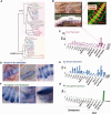
Quest for pan-neuronal genes. (A) Expression from microarray data of the four human paralogs of the ELAV genes shows that only one, ELAV4, is expressed exclusively in the neurons. (B) None of the two human paralogs of Musashi show any neural-specific expression. All microarray expression data are from BioGPS—a free gene portal for annotation and quantification of genes (Wu et al. 2009, 2013). The dataset for humans was obtained from GeneAtlas U133A, gcrma which used high-density oligonucleotide arrays to interrogate the expression of the majority of protein-encoding genes from 79 human tissues (Su et al. 2002, 2004). Protein structure for both ELAVL4 (C) and Musashi 1 (D) are from the Protein Data Bases, PDBe (Drew et al. 1981) and RCSB PDB: www.rcsb.org (Berman et al. 2000a, 2000b). ELAVL4 Protein ID: doi: 10.2210/PBD ID:1fxl (Wang and Tanaka Hall 2001) and Musashi 1 doi: 10.2210/Protein ID: 1uaw (Miyanoiri et al. 2003). Genomic organization was obtained from NCBI HomoloGene. The human ELAV genes have five to seven exons, and we used Gene ID:1996 ELAVL4 as a representative example. Musashi genes in humans have 13–14 exons, and we used Gene ID: 4440 Musashi 1 as a representative example. (This figure is available in black and white in print and in color at Integrative and Comparative Biology online.)
Fig. 2
Fig. 2
The distribution and expression of bilaterian neuronal markers in the ctenophore Pleurobrachia. (A) The genealogy of ELAV-like proteins and related members of the CELF/BRUNOL (CUGBP-like) family. The phylogenetic analysis shows that these two related protein families cluster separately. The Pleurobrachia ELAVs, 1, 3, and 4 cluster with Mnemiopsis ELAVs, and the ctenophore proteins form a separate clade basal to all other species with ELAVs. (B) Expression patterns of Pleurobrachia ELAV genes. The in situ hybridization for PbELAV3 indicates that its expression is highest in the comb plates. Insert shows immunohistochemistry for the comb plate muscles (red, in the online version) stained by phalloidin and tyrosinated α-tubulin for subepithelial neural net (green, in the online version). The staining of the muscles in the comb plates is the same location as the localization of the Pleurobrachia ELAV3 RNA in the in situ hybridization (modified from Moroz et al. 2014). (C) RNA-seq data also show highest levels of expression in the combs for the three Pleurobrachia paralogs of ELAV. (D) The in situ hybridization for the Musashi 3 RNA illustrates high levels of expression in the comb plates and non-neuronal cells around the aboral organ. (E) Quantitative expression analysis shows widespread expression of three Pleurobrachia paralogs of Musashi, including high levels of expression early in development before neuronal specification. (F) Neither in situ hybridization nor RNA-seq profiling supports differential neuronal expression of the Neuroglobin ortholog in Pleurobrachia. (G) The Neuroglobin mRNA is mainly expressed in the tentacles. See Supplementary Material for details, the accession numbers of the sequences used, RNA-seq quantification (transcript per million or TPM), and references to in situ hybridization protocols. (This figure is available in black and white in print and in color at Integrative and Comparative Biology online.)
Fig. 3
Fig. 3
Quest for canonical low-molecular-weight transmitters in ctenophores. (A) Examples of catecholamine, serotonergic, and cholinergic synthetic pathways and their phylogenetic analysis (B). A genome-wide search supported the hypothesis that the critical enzymes for each pathway are absent in Pleurobrachia (crosses X). Only phenylalanine hydroxylase (PH), which clustered phylogenetically with other PHs, was identified in both Pleurobrachia and Mnemiopsis. Neither tryptophan hydroxylase (TPH) nor tyrosine hydroxylase (TH) were detected in any ctenophore sequenced so far. We found no evidence for cholinergic signaling due to the absence of ChAT. We detected a related gene—CPT (Carnitine Palmitoyltransferase). However, all ctenophore CPT sequences cluster phylogenetically with other bilaterian CPTs, but not with ChAT (Choline Acelyltransferase). See Supplementary Material for methods and accession numbers of the sequences used in the analysis. (This figure is available in black and white in print and in color at Integrative and Comparative Biology online.)
Fig. 4
Fig. 4
Diversity of ion channels. Genome-wide complement of ion channels and receptors in representatives of five major metazoan lineages and two unicellular eukaryotes (Monosiga and Capsaspora). The receptor channels listed in the table are mostly ligand-gated or ionotropic channels with iGluR, ChRN, HTR3, GABA, and GLR highlighted as light gray. The dark gray highlights are the ENaC (SCNN1)/ASIC family; there is noticeable expansions in two ctenophore species and in Nematostella. Colored circles are examples of secondary loss in sponges and placozoans. The genomes of both Pleurobrachia and Mnemiopsis contain 131 channels/receptors compared with 61 and 49 channel-coding genes in Trichoplax and Amphimedon, respectively. It appears that there are very few ion channels or receptors that are metazoan innovations. The asterisk indicates channels identified in unicellular eukaryotes but not in the genomes of Monosiga or Capsaspora. (This figure is available in black and white in print and in color at Integrative and Comparative Biology online.)
Fig. 5
Fig. 5
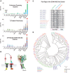
Signaling/exocytosis and ion-channel complement. (A) Numbers of the signaling/exocytosis proteins encoded in representative genomes of five basal metazoan lineages and two unicellular eukaryotes. Interestingly, the nerveless Trichoplax has more signaling/exocytosis proteins than the two ctenophore species, Pleurobrachia and Mnemiopsis. This emphasizes the fact that there may be no pan-synaptic genes. (B) Quantification of different classes of ion channels and receptors encoded in genomes from representatives of five major metazoan lineages and two unicellular eukaryotes. Here, there is a substantial difference in the numbers of ion channels and receptors in species without neurons compared with species with neural systems. (This figure is available in black and white in print and in color at Integrative and Comparative Biology online.)
Fig. 6
Fig. 6
Molecular components associated with peptidergic signaling in Pleurobrachia. (A) Quantitative (RNA-seq) expression profiles of three groups of genes supporting peptidergic signaling in Pleurobrachia: (1) proteolytic enzymes involved in prohormone processing (PC1/3, furin, and PAM); (2) 72 predicted prohormone/secretory molecules; (3) 29 putative SCNN1/ASIC-like channels and potential receptors of prohormones. Similar expression patterns were observed for all three families of genes: relatively low expression in the developmental stages and higher expression in the adult tissue with tentacles having the highest overall level of expression. The similar expression pattern suggests a possible relationship between the processing enzymes of the prohormones and their putative receptors. (B) A graphic representation of the primary structure of SCNN1/ASIC-like channels showing conserved domains and motifs. Abbreviations include CRD, TM, and conserved amino acid residues motifs. Crystal structure of ASIC1 is from the Protein Data Bases (see legend of Fig. 1 for database references) doi: 10.2210/PBD:ID 2qts (Jasti et al. 2007). (C) Alignment of conserved pore region in SCNN1/ASIC-like channels. The location and amino-acid residues for the degeneration-inducing mutation site, the amiloride-binding site, and the selective filter are indicated on the top of the alignment. The locations of transmembrane domains are listed at the bottom of the alignment. Both ctenophores’ SCNN1/ASIC-like channels contained an aspartic-acid residue (D) instead of a glycine residue (G) in the amiloride-binding site and a phenylalanine residue (F) as the x in the selectivity filter consisting of the G/SxS motif (Kellenberger et al. 2002; Kellenberger and Schild 2002, 2015). Overall, both the Pleurobrachia and Mnemiopsis ASIC-like channels have a conserved pore region. (D) The genealogical analysis of SCNN1/ASIC-like channels. All Amphimedon channels form their own clade. All human SCNN1 sequences cluster together as do the human ASIC1-4, whereas the ASIC-5 from humans clusters with Trichopax channels. The Naeglerias’ channels appear at the base of the tree. Ctenophore SCNN1/ASIC-like channels share highest identity to each other and form their own unique cluster basal to all other animals. See Supplementary Material for methods and accession numbers of the sequences used in this analysis. (This figure is available in black and white in print and in color at Integrative and Comparative Biology online.)
Fig. 7
Fig. 7
Parallel evolution of major transmitter systems in Metazoa. Five clades of the basal metazoans are shown (see Moroz et al. 2014; Whelan et al. 2015 for details of the presented phylogeny). Majority of canonical low-molecular-weight transmitters (serotonin, acetylcholine, dopamine, octopamine, noradrenaline, adrenaline, histamine, GABA) were recruited to neural signaling in the bilaterian–cnidarian ancestor, while ctenophores had independently developed a diversity of intercellular messengers (including signaling peptides) for neural communications. l-Glutamate might also be independently recruited for neural signaling in ctenophores and bilaterian/cnidarian clades. Sponges (Porifera) and placozoans represent lineages with the primary absence of neural systems. Two lighting signs indicate origins of neurons, synapses, muscles, and mesoderm in ctenophores and bilaterian/cnidarian clades, respectively. (This figure is available in black and white in print and in color at Integrative and Comparative Biology online.)

References

    1. Abascal F, Zardoya R. 2013. Evolutionary analyses of gap junction protein families. Biochim Biophys Acta 1828:4–14. - PubMed
    1. Anctil M. 1985. Cholinergic and monoaminergic mechanisms associated with control of bioluminescence in the ctenophore Mnemiopsis leidyi. J Exp Biol 119:225–38.
    1. Anctil M. 2009. Chemical transmission in the sea anemone Nematostella vectensis: A genomic perspective. Comp Biochem Physiol Part D Genomics Proteomics 4:268–89. - PubMed
    1. Anne C, Gasnier B. 2014. Vesicular neurotransmitter transporters: Mechanistic aspects. Curr Top Membr 73:149–74. - PubMed
    1. Bang ML, Owczarek S. 2013. A matter of balance: Role of neurexin and neuroligin at the synapse. Neurochem Res 38:1174–89. - PubMed

Publication types

Substances