. 2015 Nov 2;25(21):2830-2838.
doi: 10.1016/j.cub.2015.09.026. Epub 2015 Oct 8.

Catalytic strand separation by RECQ1 is required for RPA-mediated response to replication stress

Affiliations

Catalytic strand separation by RECQ1 is required for RPA-mediated response to replication stress

Taraswi Banerjee et al. Curr Biol. .

Abstract

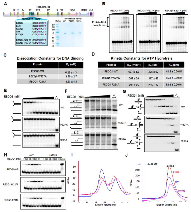
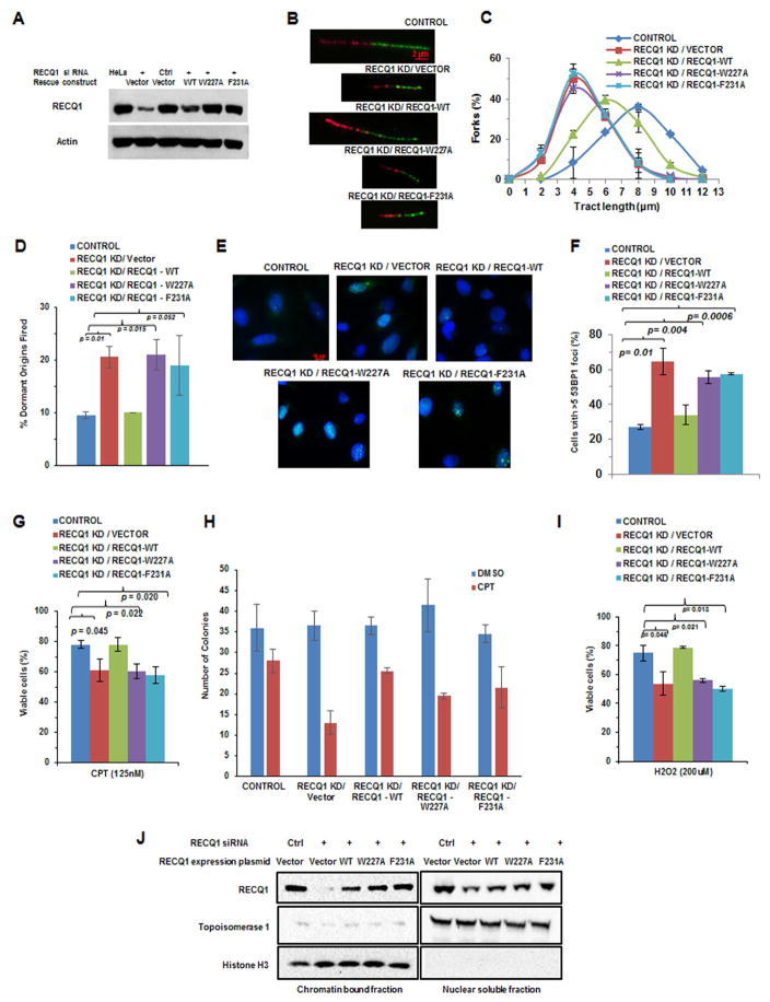
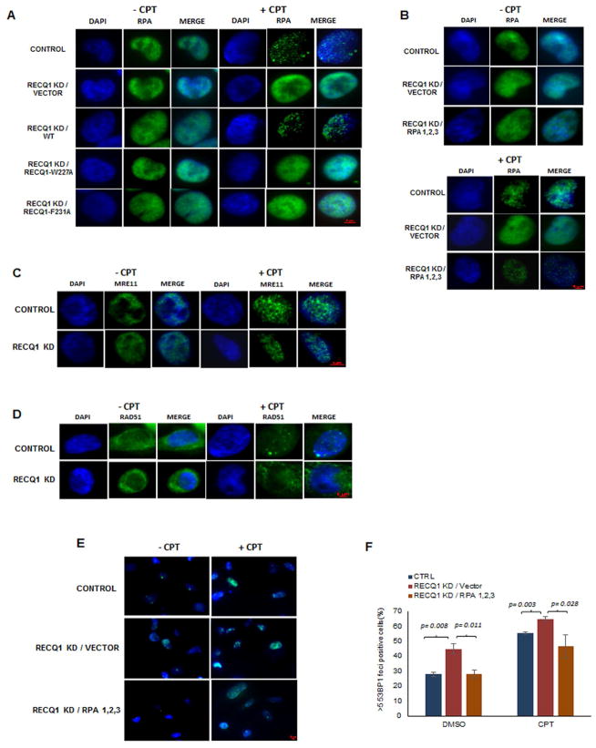
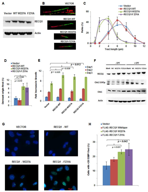
Three (BLM, WRN, and RECQ4) of the five human RecQ helicases are linked to genetic disorders characterized by genomic instability, cancer, and accelerated aging [1]. RECQ1, the first human RecQ helicase discovered [2-4] and the most abundant [5], was recently implicated in breast cancer [6, 7]. RECQ1 is an ATP-dependent DNA-unwinding enzyme (helicase) [8, 9] with roles in replication [10-12] and DNA repair [13-16]. RECQ1 is highly expressed in various tumors and cancer cell lines (for review, see [17]), and its suppression reduces cancer cell proliferation [14], suggesting a target for anti-cancer drugs. RECQ1's assembly state plays a critical role in modulating its helicase, branch migration (BM), or strand annealing [18, 19]. The crystal structure of truncated RECQ1 [20, 21] resembles that of E. coli RecQ [22] with two RecA-like domains, a RecQ-specific zinc-binding domain and a winged-helix domain, the latter implicated in DNA strand separation and oligomer formation. In addition, a conserved aromatic loop (AL) is important for DNA unwinding by bacterial RecQ [23, 24] and truncated RECQ1 helicases [21]. To better understand the roles of RECQ1, two AL mutants (W227A and F231A) in full-length RECQ1 were characterized biochemically and genetically. The RECQ1 mutants were defective in helicase or BM but retained DNA binding, oligomerization, ATPase, and strand annealing. RECQ1-depleted HeLa cells expressing either AL mutant displayed reduced replication tract length, elevated dormant origin firing, and increased double-strand breaks that could be suppressed by exogenously expressed replication protein A (RPA). Thus, RECQ1 governs RPA's availability in order to maintain normal replication dynamics, suppress DNA damage, and preserve genome homeostasis.


Figures

Figure 1
Figure 1. Biochemical analysis of purified recombinant human RECQ1 proteins
(A) Schematic of human RECQ1 showing conserved domains and helicase core signature motifs. The conserved aromatic-rich sequence between motifs II and III is shown with a sequence alignment of the five human RecQ helicases. The two invariant aromatic amino acids and corresponding amino acid substitutions in RECQ1 are indicated in pink and blue, respectively (left). Coomassie-stained SDS polyacrylamide gel showing recombinant human RECQ1 proteins purified from insect cells (right). (B) Autoradiogram showing representative gel from EMSA experiments with RECQ1 proteins (0.15, 0.31, 0.62, 1.25, 2.5, 5, 10, and 20 nM monomer) incubated with a forked 19 bp duplex DNA substrate. (C) Apparent dissociation constant values (Kd) estimating binding affinities of RECQ1 proteins for the forked duplex substrate. (D) Kinetic constants for ATP hydrolysis by RECQ1 proteins. (E) Autoradiogram showing representative native polyacrylamide gel from helicase assays with RECQ1 proteins (0.15, 0.31, 0.62, 1.25, 2.5, 5, 10, 20 and 40 nM monomer) and the 19 bp forked duplex DNA substrate. (F) Autoradiogram showing representative native polyacrylamide gel from BM assays with RECQ1 proteins (25, 50,100, and 200 nM monomer) and mobile three-stranded D-loop DNA substrate. (G) Autoradiogram showing representative native polyacrylamide gel from BM assays with RECQ1 proteins (0.62, 1.25, 2.5, 5, 10, and 20 nM monomer) and the four-stranded HJ DNA substrate. (H) Autoradiogram showing representative native polyacrylamide gel from strand annealing assays with RECQ1 proteins (1.25, 2.5, 5, 10, 20, and 40 nM monomer) and complementary single-stranded oligonucleotides in the absence or presence of 1 mM ATPγS as indicated. (I, J) Size exclusion chromatogram showing distribution of purified RECQ1 proteins incubated in the absence (I) or presence (J) of 1 mM ATP.
Figure 2
Figure 2. Perturbed replication and genomic instability of human cells expressing RECQ1-W227A or RECQ1-F231A aromatic loop mutant proteins
(A) Representative Western blot from lysates of HeLa cells transfected with control siRNA or siRNA against 3′-UTR of RECQ1 and an expression construct encoding the indicated RECQ1 protein. Actin serves as a loading control. (B) RECQ1-depleted cells (RECQ1 KD) exogenously expressing RECQ1-WT, RECQ1-W227A, or RECQ1-F231A protein were labeled with CldU or IdU indicated by red and green tracts, respectively. Representative DNA fiber tracts from microfluidic-assisted tract analysis are shown. (C) Statistical analysis of percentage replication forks with the indicated tract lengths (μm). At least 100 tracts were analyzed for each cell line. (D) Percentage of dormant origins fired for indicated cell lines. (E) Representative 53BP1 fluorescence staining images of indicated cell lines. (F) Quantitative analysis of immunofluorescence data as shown in (E). Percentage of cells with >5 53BP1 foci is shown. (G) Western blot showing the relative binding of RECQ1 proteins to chromatin isolated from CPT-treated cells. (H) The indicated cell line was exposed to CPT (125 nM, 16 hr) and percent viable cells determined by WST-1 assay. (I) Indicated cell lines were exposed to 100 nM CPT for 1 hr and monitored after 13 days for colony formation. (J) Cell lines were exposed to H2O2 (200 μM, 30 min) and percent viable cells was determined by WST-1 assay.
Figure 3
Figure 3. DNA damage in HeLa cells expressing catalytically defective RECQ1 is a function of RPA exhaustion
(A) Representative immunofluorescence images of RPA staining in indicated HeLa cell lines treated with either DMSO or 200 nM CPT for 30 min. Representative immunofluorescence images of RPA (B), MRE11 (C), RAD51 (D), and 53BP1 (E) staining in indicated HeLa cell lines treated with either DMSO or 200 nM CPT for 30 min. (F) Quantitative analysis of 53B1 staining in cells represented in E.
Figure 4
Figure 4. Dominant negative effects exerted by expression of RECQ1 aromatic loop mutant proteins
(A) Representative Western blot from lysates of HeLa cells transfected with an expression construct encoding the indicated RECQ1 protein. Actin serves as a loading control. (B) HeLa cells exogenously expressing RECQ1-WT, RECQ1-W227A, or RECQ1-F231A protein were labeled with CldU or IdU indicated by red and green tracts, respectively. Representative DNA fiber tracts from microfluidic-assisted tract analysis are shown for each of the cell lines. (C) Statistical analysis of percentage replication forks with the indicated tract lengths (μm). At least 100 tracts were analyzed for each cell line. (D) Percentage of dormant origins fired for the indicated cell lines. (E) Graph comparing the growth pattern of HeLa cells over-expressing exogenous wild-type or either of the two RECQ1 AL mutant proteins over a period of 3 days. (F) Western blot showing Chk1 S345 phosphorylation in untreated and CPT-treated (2 μM, 1 hr) HeLa cells transfected with blank vector, wild-type RECQ1 or either AL mutant RECQ1 expressing construct. CPT-treated RECQ1-W227A and RECQ1-F231A transfected cells displayed 47% and 79% less phosphorylated Chk1S345, respectively, compared to CPT-treated cells that were transfected with blank vector, as normalized against total Chk1. (G) Representative fluorescence images from 53BP1 staining of indicated cell lines. (H) Quantitative analysis of immunofluorescence data as shown in (G). Percentage of cells with >20 53BP1 foci is shown.

Similar articles

Cited by

References

    1. Brosh RM., Jr DNA helicases involved in DNA repair and their roles in cancer. Nat Rev Cancer. 2013;13:542–548. - PMC - PubMed
    1. Seki M, Yanagisawa J, Kohda T, Sonoyama T, Ui M, Enomoto T. Purification of two DNA-dependent adenosinetriphosphatases having DNA helicase activity from HeLa cells and comparison of the properties of the two enzymes. J Biochem. 1994;115:523–531. - PubMed
    1. Puranam KL, Kennington E, Sait SN, Shows TB, Rochelle JM, Seldin MF, Blackshear PJ. Chromosomal localization of the gene encoding the human DNA helicase RECQL and its mouse homologue. Genomics. 1995;26:595–598. - PubMed
    1. Puranam KL, Blackshear PJ. Cloning and characterization of RECQL, a potential human homologue of the Escherichia coli DNA helicase RecQ. J Biol Chem. 1994;269:29838–29845. - PubMed
    1. Kawabe T, Tsuyama N, Kitao S, Nishikawa K, Shimamoto A, Shiratori M, Matsumoto T, Anno K, Sato T, Mitsui Y, et al. Differential regulation of human RecQ family helicases in cell transformation and cell cycle. Oncogene. 2000;19:4764–4772. - PubMed

Publication types