. 2015 Oct 15;60(2):231-41.
doi: 10.1016/j.molcel.2015.09.006. Epub 2015 Oct 8.

Residue-by-Residue View of In Vitro FUS Granules that Bind the C-Terminal Domain of RNA Polymerase II

Affiliations

Residue-by-Residue View of In Vitro FUS Granules that Bind the C-Terminal Domain of RNA Polymerase II

Kathleen A Burke et al. Mol Cell. .

Abstract

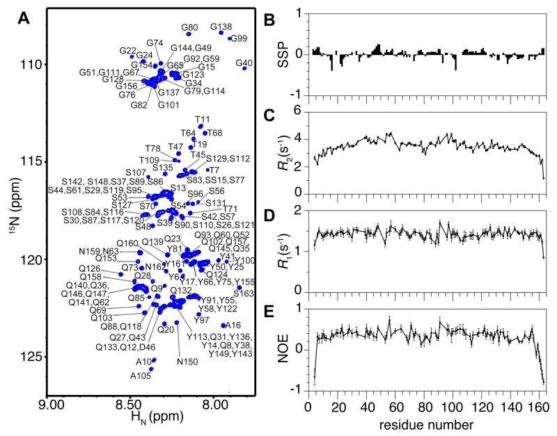
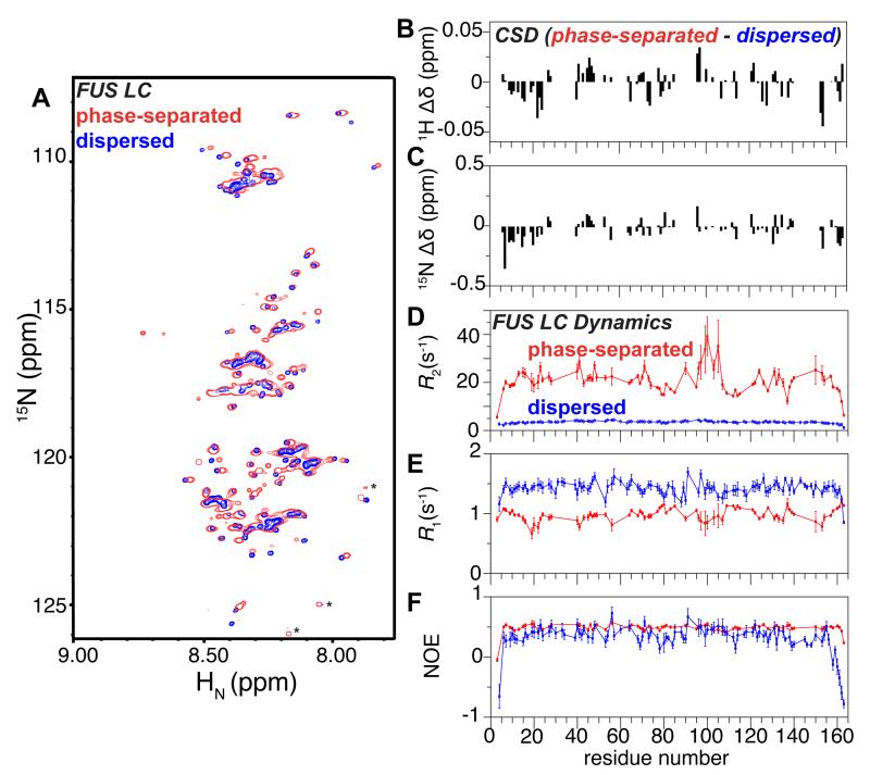
Phase-separated states of proteins underlie ribonucleoprotein (RNP) granules and nuclear RNA-binding protein assemblies that may nucleate protein inclusions associated with neurodegenerative diseases. We report that the N-terminal low-complexity domain of the RNA-binding protein Fused in Sarcoma (FUS LC) is structurally disordered and forms a liquid-like phase-separated state resembling RNP granules. This state directly binds the C-terminal domain of RNA polymerase II. Phase-separated FUS lacks static structures as probed by fluorescence microscopy, indicating they are distinct from both protein inclusions and hydrogels. We use solution nuclear magnetic resonance spectroscopy to directly probe the dynamic architecture within FUS liquid phase-separated assemblies. Importantly, we find that FUS LC retains disordered secondary structure even in the liquid phase-separated state. Therefore, we propose that disordered protein granules, even those made of aggregation-prone prion-like domains, are dynamic and disordered molecular assemblies with transiently formed protein-protein contacts.


Figures

Figure 1
Figure 1
The FUS low complexity (LC, residues 1-163) domain is disordered as a monomer. (A) NMR spectrum (1H-15N heteronuclear single quantum coherence, HSQC) of the backbone amide region of FUS LC has a narrow chemical shift dispersion indicative of a disordered protein. Residue numbers are labeled (black). (B) Residue-specific secondary structure propensity (SSP) score of FUS LC indicate lack of local secondary structure formation. R2, R1, and (1H)-15N nuclear Overhauser effect (NOE) values (C, D, E, respectively) for the dispersed protein are consistent with disorder across the entire domain. Data are represented as mean +/− st dev. See also Figure S1.
Figure 2
Figure 2
The low complexity domain of FUS stabilizes phase separation that recruits the RNA polymerase II C-terminal domain. (A) Differential interference contrast (DIC) microscopy shows that the isolated FUS LC phase-separates at room temperature at high concentration (left) but not at low concentration (right). (B) 10 μM FUS full-length protein phase-separates only when liberated from an N-terminal maltose binding protein (MBP) fusion by addition of TEV protease (left) but not when incubated with buffer (right). (C) Phase separation of FUS LC as measured by optical density at 600 nm as a function of temperature and protein concentration, starting at 20 °C down to 0 °C. By increasing the temperature of the same samples back up to 20 °C, the phase-separated state dissipates, indicating the assembly is predominantly reversible. FUS LC phase separation is enhanced at increasing salt concentrations (left to right). (D) Phase separation of 5 μM full-length FUS after cleavage from a solubilizing MBP fusion (MBP-FUS) is unaffected by increasing NaCl concentration from 50 to 150 mM but reduced in 300 mM NaCl. Data are represented as triplicate mean +/− st dev. See also Figure S2.
Figure 3
Figure 3
Intermolecular interactions in phase separated FUS. (A) Phase separation of 5 μM FUS (after cleaving from MBP fusion by TEV protease, see Figure 2B) is enhanced by addition of torula yeast RNA up to a weight ratio of 0.4:1. (B) Fusions of GFP to the 26 degenerate heptad of the C-terminal domain of RNA polymerase II (GFP-CTD) localize to phase separated states of FUS LC. Data are represented as mean +/− st dev. See also Figure S3.
Figure 4
Figure 4
Rapid diffusion and turnover of FUS LC droplet contents. Example differential interference contrast and fluorescence images (left panels) and FRAP timecourses (right) of partial droplet photobleaching experiments used to measure diffusion constants within the droplets of FUS LC + FUS LC-Alexa (A) and FUS LC + GFP-CTD (B). Half times for the selected timecourse are labeled in the recovery curves. Example differential interference contrast and fluorescence images (left panels) and FRAP timecourses (right) of full droplet photobleaching experiments used to measure rate of exchange between droplet and dispersed states of FUS LC + FUS LC-Alexa (C) and FUS LC + GFP-CTD (D). See also Figure S4.
Figure 5
Figure 5
Phase separated FUS LC remains disordered and dynamic. (A) 1H 15N HSQC spectrum of the phase separated FUS LC is highly similar to the monomer in the dispersed state solution, suggesting a similar overall conformation. Asterisks denote presumed C-termini of small quantities of impurities or truncations where favorable relaxation properties amplify their signal compared to FUS LC resonances. 1H (B) and 15N (C) chemical shift differences between FUS LC in the phase-separated and dispersed states indicate that small chemical shift differences are distributed across the chain. R2, R1, and (1H)-15N nuclear Overhauser effect (NOE) values (D, E, F, respectively) for phase separated FUS LC (red) are consistent with slowed motion compared to the dispersed phase (blue, repeated from Figure 1) but demonstrate that FUS LC retains reorientational mobility. Data are represented as mean +/− st dev. See also Figure S5.
Figure 6
Figure 6
NMR resonance intensities arising from phase separated FUS LC are highly temperature dependent. One dimensional 1H (A) and 1H 15N HSQC (B) experiments show decreased signal intensity and increased line broadening as temperature decreases.

Similar articles

Cited by

References

    1. Banjade S, Rosen MK. Phase transitions of multivalent proteins can promote clustering of membrane receptors. Elife. 2014;3 - PMC - PubMed
    1. Belzil VV, Valdmanis PN, Dion PA, Daoud H, Kabashi E, Noreau A, Gauthier J, Hince P, Desjarlais A, Bouchard JP, et al. Mutations in FUS cause FALS and SALS in French and French Canadian populations. Neurology. 2009;73:1176–1179. - PMC - PubMed
    1. Bentmann E, Neumann M, Tahirovic S, Rodde R, Dormann D, Haass C. Requirements for stress granule recruitment of fused in sarcoma (FUS) and TAR DNA-binding protein of 43 kDa (TDP-43) The Journal of biological chemistry. 2012;287:23079–23094. - PMC - PubMed
    1. Bodner CR, Dobson CM, Bax A. Multiple tight phospholipid-binding modes of alpha-synuclein revealed by solution NMR spectroscopy. Journal of molecular biology. 2009;390:775–790. - PMC - PubMed
    1. Brangwynne CP. Phase transitions and size scaling of membrane-less organelles. J Cell Biol. 2013;203:875–881. - PMC - PubMed

Publication types

MeSH terms