Review
. 2017 Aug:155:96-119.
doi: 10.1016/j.pneurobio.2015.09.012. Epub 2015 Oct 9.

Interactions of iron, dopamine and neuromelanin pathways in brain aging and Parkinson's disease

Affiliations
Review

Interactions of iron, dopamine and neuromelanin pathways in brain aging and Parkinson's disease

Fabio A Zucca et al. Prog Neurobiol. 2017 Aug.

Abstract

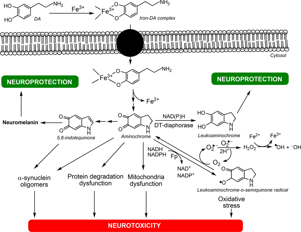
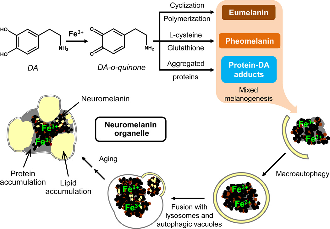
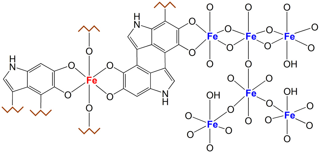
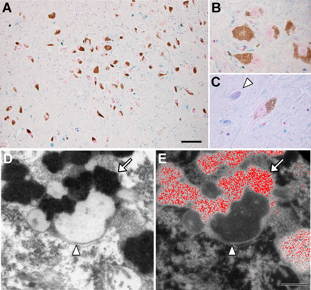
There are several interrelated mechanisms involving iron, dopamine, and neuromelanin in neurons. Neuromelanin accumulates during aging and is the catecholamine-derived pigment of the dopamine neurons of the substantia nigra and norepinephrine neurons of the locus coeruleus, the two neuronal populations most targeted in Parkinson's disease. Many cellular redox reactions rely on iron, however an altered distribution of reactive iron is cytotoxic. In fact, increased levels of iron in the brain of Parkinson's disease patients are present. Dopamine accumulation can induce neuronal death; however, excess dopamine can be removed by converting it into a stable compound like neuromelanin, and this process rescues the cell. Interestingly, the main iron compound in dopamine and norepinephrine neurons is the neuromelanin-iron complex, since neuromelanin is an effective metal chelator. Neuromelanin serves to trap iron and provide neuronal protection from oxidative stress. This equilibrium between iron, dopamine, and neuromelanin is crucial for cell homeostasis and in some cellular circumstances can be disrupted. Indeed, when neuromelanin-containing organelles accumulate high load of toxins and iron during aging a neurodegenerative process can be triggered. In addition, neuromelanin released by degenerating neurons activates microglia and the latter cause neurons death with further release of neuromelanin, then starting a self-propelling mechanism of neuroinflammation and neurodegeneration. Considering the above issues, age-related accumulation of neuromelanin in dopamine neurons shows an interesting link between aging and neurodegeneration.

Keywords: Dopamine; Human neuromelanin; Iron; Melanin; Parkinson's disease.


Figures

Fig. 1
Fig. 1. The neurotoxicity of iron-DA complex
DA forms complex with ferric iron (Fe3+) that is taken up into the cell by DA transporters. Iron-DA complex undergoes a redox reaction where ferric iron is reduced to ferrous iron (Fe2+) and DA oxidizes to aminochrome. Then aminochrome can polymerize to NM via 5,6-indolequinone affording neuroprotection, or can be two-electron reduced by DT-diaphorase to leukoaminochrome, preventing aminochrome-induced neurotoxic reactions such as: i) α-synuclein aggregation to neurotoxic oligomers; ii) protein degradation dysfunction; iii) mitochondria dysfunction; iv) oxidative stress caused by one-electron reduction of aminochrome to leukoaminochrome-o-semiquinone radical catalyzed by flavoenzymes (Fp). Leukoaminochrome-o-semiquinone radical can auto-oxidize to aminochrome by reducing dioxygen (O2) to superoxide radical anion (O2•−): then the dismutation of two superoxide radicals regenerates O2 and yields hydrogen peroxide (H2O2), which in presence of ferrous iron forms the hydroxyl radical (OH) by Fenton's reaction, leading to high oxidative stress.
Fig. 2
Fig. 2. DA oxidation to o-quinones
DA oxidizes to DA-o-quinone that can form adducts with parkin, resulting in the inactivation of the proteasomal system. DA-o-quinone can form adducts also with mitochondrial complex I, III and V inducing mitochondrial dysfunction. DA-o-quinone can undergo intramolecular cyclization to aminochrome, which can: i) inactivate the proteasome; ii) inactivate mitochondria complex I inducing mitochondrial dysfunction; iii) be one-electron reduced by flavoenzymes to leukoaminochrome-o-semiquinone radical, which auto-oxidizes immediately to aminochrome generating oxidative stress; iv) inactivate the vacuolar ATPase proton pump of lysosomes inducing lysosomal dysfunction; v) induce aggregation of α- and β-tubulin preventing the formation of microtubules required for the fusion of autophagic vacuoles with lysosomes, thus generating autophagic dysfunction; vi) induce α-synuclein aggregation to neurotoxic oligomers. The neurotoxic reactions of aminochrome can be prevented by aminochrome polymerization to NM (via rearrangement to 5,6-dihydroxindole and then to 5,6-indolequinone) or two-electron reduction, catalyzed by DT-diaphorase, of aminochrome to leukoaminochrome that probably can undergo rearrangement to 5,6-dihydroxyindole, the precursor of 5,6-indolequinone, which can be involved as well in NM synthesis.
Fig. 3
Fig. 3. Possible mechanisms for the synthesis of NM pigment and for the formation of NM-containing organelles
Excess of DA present in the cytosol can be oxidized to DA-o-quinone by ferric iron in a catalytic reaction. In the formation of NM pigment, DA-o-quinone can undergo three different pathways: i) cyclization, further oxidation and polymerization to give eumelanin; ii) reaction with L-cysteine or glutathione to give cysteinyl-DA compounds then oxidized to pheomelanin; and iii) conjugation with protein residues to give DA-protein adducts. The latter two reactions seem to be faster and lead to the formation of a protein-pheomelanin core, which is then coated by eumelanin, according to the mixed melanogenesis model. Iron(III) is incorporated into the melanic portion of the forming NM pigment. The resulting undegradable and insoluble pigment is taken into autophagic vacuoles that fuse with lysosomes and other autophagic vacuoles containing lipids, proteins, etc., leading to the formation of NM-containing organelles. These double membrane bounded organelles contain NM pigment along with its components, abundant lipid bodies, and protein matrix. This process continues during the entire neuron life and results in the accumulation of NM-containing organelles with aging. This scheme is modified from Zucca and colleagues (2014) by permission of Springer Publishing.
Fig. 4
Fig. 4. Iron centers in NM pigment
The figure shows the two types of iron centers present in the NM pigment. The multinuclear iron cluster, similarly to ferritin, contains iron(III) ions (blue colored in the figure) coupled by oxy-hydroxy bridges and surrounded by catechol groups of NM. In this site, iron is probably stored with high affinity and maintained in a redox inactive state, and is principally detected by Mössbauer spectroscopy. The other iron site consists of mononuclear centers, where iron (red colored) is coordinated by oxygen atoms of catechols moieties, and possibly by hydroxo groups. This could be a low affinity binding site occupied only in case of iron overload, when the high affinity centers are saturated; in iron overload condition as occurring in PD, the mononuclear iron could be still redox reactive and catalyze the production of toxic species, for example via the Fenton’s reaction. Iron in this site is principally detected by EPR spectroscopy.
Fig. 5
Fig. 5. Iron deposits in healthy aged SN
(A, B, C) Histochemistry with modified Perls’ staining for detection of reactive ferric iron deposits (for details see Zecca et al., 2004b) in normal human SN tissue sections (88 years old). Many iron deposits are present in glial cells and are stained in blue: the overview (A, scale bar = 100 µm) shows that blue iron deposits are abundant in the whole SN parenchyma, while are absent in NM-containing neurons (brown-colored for the presence of NM pigment in the cytoplasm), as confirmed at higher magnification showing a group of NM-containing neurons (B). However NM-free neurons of the SN contain large amounts of iron deposits (arrowhead in C). Panels A, B, C are modified from Zucca and colleagues (2011) by permission of Royal Society of Chemistry Publishing. (D, E) NM-containing organelles of normal human SN (89 years old) observed using electron imaging. In the left panel (D), transmission electron microscopy shows the classical structure of NM-containing organelles present in SN neurons: these organelles contain large amount of dark NM pigment (arrow) closely associated with lipid bodies (arrowhead) as previously reported (Sulzer et al., 2008; Zecca et al., 2008b). The elemental iron distribution map obtained by electron spectroscopic imaging (E, scale bar = 500 nm) clearly reveals that large amounts of iron deposits are localized inside the NM pigment of the organelle (iron element is shown in red color). This unpublished finding strongly confirms the ability of NM pigment to scavenge iron forming stable NM-iron complexes. Electron spectroscopic imaging were performed by using a LEO 912AB electron microscope as described by Pezzati and colleagues (1997). For tissue treatments and ethics policies refer to: Zecca et al., 2008b; Engelen et al., 2012.
Fig. 6
Fig. 6. Iron deposits in different regions of healthy aged brain
(A, B, C) Putamen of 71 years old subject. (A, scale bar = 100 µm). Reactive ferric iron deposits visualized by Perls’ histochemistry (for details see Zecca et al., 2004b) are clearly abundant in the whole parenchyma and principally localized in glial cells (diffuse blue staining) and fibers (asterisk in C). Iron deposits are clearly present in the cytoplasm of two neurons not containing NM (arrowhead in A), as confirmed at higher magnification (arrowhead in B). NM-containing neurons of putamen (brown-colored for the presence of NM pigment) do not show blue staining in their cytoplasm (arrowheads in C). (D, E, F) Globus pallidus of 71 years old subject. (D, scale bar = 100 µm). The iron distribution in the globus pallidus is similar to that observed in the putamen. Iron deposits are abundant and mainly present in glial cells (diffuse blue staining) and in fibers (asterisks in D and E). Cytoplasm of neurons not containing NM (arrowhead in E) is clearly positive for iron staining, while NM-containing neuron (slightly brown-colored for the presence of NM pigment) apparently does not contain iron deposits (arrowhead in F). (G, H, I) Premotor cortex of 72 years old subject. Iron deposits in the premotor cortex are fewer if compared to other previous brain areas (G, scale bar = 100 µm). Scarce iron deposits are mainly present in glial cells (arrows indicate some deposits in G) and are completely absent in both neurons without visible NM (arrowheads in H) and in NM-containing neurons (arrowhead in I, indicating a neuron with weak brown-colored pigmentation due to the presence of NM). (J, K, L) Cerebellum of 80 years old subject. (J, scale bar = 100 µm). Iron deposits are scattered in the different layers of the cerebellar architecture (J): the arrow indicates iron in the molecular layer, arrowhead in the granular layer and asterisk in the white matter. However the most intense staining is localized in the white matter of the cerebellum. Panels K and L show at higher magnification the Purkinje cells layer: iron staining is sometimes observed in the middle layer between molecular and granular layers (arrow in K), while Purkinje cells apparently do not contain iron deposits in their cytoplasm (arrowhead in L). The presence of NM pigments has been recently established by means of different techniques in neurons of putamen, globus pallidus, premotor cortex, cerebellum, and other human brain areas (Engelen et al., 2012; Zecca et al., 2008b). However, the concentration of NM pigment in these areas is lower than that observed in SN and LC (Zecca et al., 2004b, 2008b). For tissue treatments and ethics policies refer to: Zecca et al., 2008b; Engelen et al., 2012.

Similar articles

Cited by

References

    1. Abbas N, Lücking CB, Ricard S, Dürr A, Bonifati V, De Michele G, Bouley S, Vaughan JR, Gasser T, Marconi R, Broussolle E, Brefel-Courbon C, Harhangi BS, Oostra BA, Fabrizio E, Böhme GA, Pradier L, Wood NW, Filla A, Meco G, Denefle P, Agid Y, Brice A. A wide variety of mutations in the parkin gene are responsible for autosomal recessive parkinsonism in Europe. French Parkinson's Disease Genetics Study Group and the European Consortium on Genetic Susceptibility in Parkinson's Disease. Hum. Mol. Genet. 1999;8:567–574. - PubMed
    1. Abbott NJ, Rönnbäck L, Hansson E. Astrocyte-endothelial interactions at the blood-brain barrier. Nat. Rev. Neurosci. 2006;7:41–53. - PubMed
    1. Abbott NJ, Patabendige AA, Dolman DE, Yusof SR, Begley DJ. Structure and function of the blood-brain barrier. Neurobiol. Dis. 2010;37:13–25. - PubMed
    1. Abdel-Malek Z, Kadekaro AL. Human Pigmentation: Its regulation by Ultraviolet Light and by Endocrine, Paracrine and Autocrine Factors. In: Nordlund JJ, Boissy RE, Hearing VJ, King RA, Oetting WS, Ortonne J-P, editors. The Pigmentary System: Physiology and Pathophysiology. Second edition. Malden, MA, USA: Blackwell Publishing; 2006. pp. 410–420.
    1. Agrup G, Hansson C, Rorsman H, Rosengren E. The effect of cysteine on oxidation of tyrosine, dopa, and cysteinyldopas. Arch. Dermatol. Res. 1982;272:103–115. - PubMed