. 2015 Dec;171(5):862-71.
doi: 10.1111/bjh.13778. Epub 2015 Oct 12.

The human Kell blood group binds the erythroid 4.1R protein: new insights into the 4.1R-dependent red cell membrane complex

Affiliations

The human Kell blood group binds the erythroid 4.1R protein: new insights into the 4.1R-dependent red cell membrane complex

Slim Azouzi et al. Br J Haematol. 2015 Dec.

Abstract

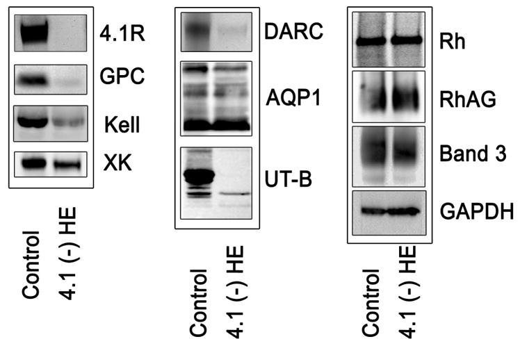
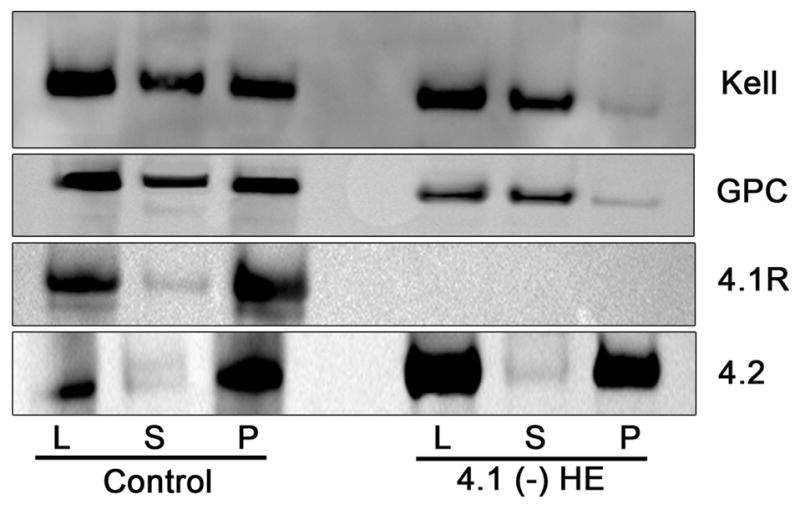
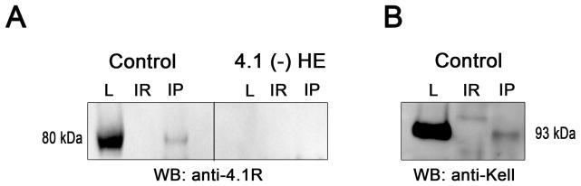
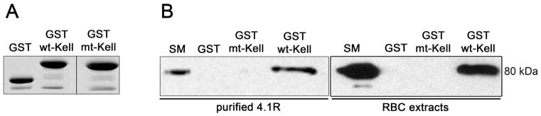
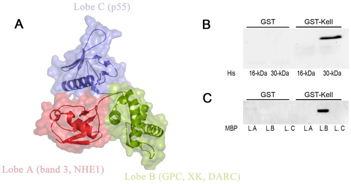
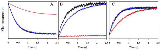
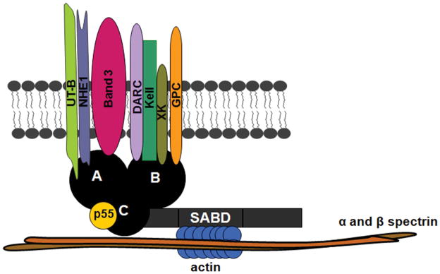
Protein 4.1R plays an important role in maintaining the mechanical properties of the erythrocyte membrane. We analysed the expression of Kell blood group protein in erythrocytes from a patient with hereditary elliptocytosis associated with complete 4.1R deficiency (4.1(-) HE). Flow cytometry and Western blot analyses revealed a severe reduction of Kell. In vitro pull down and co-immunoprecipitation experiments from erythrocyte membranes showed a direct interaction between Kell and 4.1R. Using different recombinant domains of 4.1R and the cytoplasmic domain of Kell, we demonstrated that the R(46) R motif in the juxta-membrane region of Kell binds to lobe B of the 4.1R FERM domain. We also observed that 4.1R deficiency is associated with a reduction of XK and DARC (also termed ACKR1) proteins, the absence of the glycosylated form of the urea transporter B and a slight decrease of band 3. The functional alteration of the 4.1(-) HE erythrocyte membranes was also determined by measuring various transport activities. We documented a slower rate of HCO3 (-) /Cl(-) exchange, but normal water and ammonia transport across erythrocyte membrane in the absence of 4.1. These findings provide novel insights into the structural organization of blood group antigen proteins into the 4.1R complex of the human red cell membrane.

Keywords: 4.1R; Kell protein; blood group antigens; erythrocyte membrane; macromolecular complex.


Figures

Fig. 1
Fig. 1
Western blotting analysis of membranes proteins from normal and 4.1 (−) HE erythrocytes. Membrane proteins from whole ghost lysates separated by SDS-PAGE were transferred to a nitrocellulose membrane and incubated with relevant primary antibodies against the indicated proteins. The following antibodies were used: anti 4.1R (PAb); anti-GPC (MAb 1F6); anti-Kell (MAb 5A11); anti XK (rabbit PAb); anti DARC (MAb NaM185-2C3); anti AQP1 (PAb AB3065); anti Rh (PAb MPC8); anti UT-B (rabbit PAb); anti RhAG (MAb LA18.18); anti band 3 (rabbit PAb); anti GAPDH (MAb). GADPH was used as loading control to show that similar amounts of membrane proteins were present on each lane. All experiments were repeated at least three times. HE, hereditary elliptocytosis; MAb, monoclonal antibody; PAb, polyclonal antibody.
Fig. 2
Fig. 2
Comparison of Triton X-100 extractability of the Kell protein from normal and 4.1 (−) HE erythrocytes. Erythrocyte ghosts (L) were extracted with 1% Triton-X-100, and the soluble (S) and insoluble (Pellet, P) fractions were analysed by immunoblotting. Immunostaining of Kell, GPC and 4.1R proteins was performed as described in the legend to Figure 1. Anti- 4.2 protein, which is not decreased in 4.1 (−) HE, was used to control proteins extraction for each normal and variant sample.
Fig. 3
Fig. 3
Co-immunoprecipitation of 4.1R and Kell. Erythrocytes were lysed (L) and extracts were subjected to immunoprecipitation with specific antibody (IP) or mouse irrelevant antibody (IR, anti-VCAM1 antibody). (A) Immunoprecipitation with anti-Kell monoclonal antibodies. (B) Immunoprecipitation with anti-4.1 polyclonal antibodies. Presence of the Kell protein or 4.1R protein was studied by Western blot (WB) in lysates (L) and IP eluates (IR and IP).
Fig. 4
Fig. 4
Association of 4.1R with the cytoplasmic domain of Kell. (A) Coomassie blue staining of GST, GST-wt-Kell and GST-mt-Kell aliquots before the pull down assays (12.5% acrylamide gel). Vertical line indicates grouping of images from two different gels. (B) GST- pull down was performed using purified protein 4.1R or low ionic strength red cell membrane extracts with GST, GST-wt-Kell and GST-mt-Kell. Eluted proteins were revealed with anti-4.1R polyclonal antibodies. SM: starting material; RBC, red blood cell.
Fig. 5
Fig. 5
Direct binding of Kell protein to FERM binding domain of 4.1R. The binding of 4.1R functional domains to cytoplasmic tails of Kell, was assessed by pull down assay. (A) Crystal structure of the FERM domain (30 kDa domain) of 4.1R (PDB accession ID: 1gg3). The FERM domain consists of three lobes (A, B and C), which bind to different transmembrane proteins. The binding of His-tagged domains (16 and 30 kDa domains) of 4.1R (B) and MBP-tagged subdomains of FERM domain (C) to the cytoplasmic domains of Kell, was detected by using anti- His and anti-MBP antibodies, respectively. L.A, lobe A; L.B, lobe B; L.C lobe C.
Fig. 6
Fig. 6
Comparison of water, bicarbonate and ammonia transport in ghosts from erythrocyte variants. (A) Time courses of fluorescence quenching corresponding to osmotic water permeability of ghost from control (black), 4.1Rnull (blue) and AQP1null erythrocytes (red). Ghosts were resealed in presence of 8 mM 6-carboxyfluorescein and submitted to an osmotic gradient (150 mosm mannitol/kg H2O) at 15°C in the stopped-flow spectrofluorometer. (B) Time courses of intracellular pH increase in ghosts corresponding to HCO3/Cl exchange. Ghosts derived from control (black) and 4.1Rnull (blue) erythrocytes resealed in the presence of pyranine, a fluorescent pH-sensitive probe, and bovine carbonic anhydrase at 2 mg/ml, were rapidly mixed with an equal volume of buffer containing KHCO3, generating inwardly-directed 50 MEq HCO3/CO2 and outwardly-directed 50 MEq Cl gradients. Ghost from control was incubated with the anion exchanger inhibitor DIDS (30 μM) for 20 min (red curve). (C) Time course of intracellular pH increase corresponding to ammonia transport in ghosts from control (black), 4.1Rnull (blue) and RhAGnull erythrocytes (red). Ghost resealed in the presence of pyranine were submitted to 10 mM inwardly-directed gradient of ammonia at 15°C followed by stopped-flow analysis. All experiments were repeated twice. Each kinetic corresponds to three averaged time courses of fluorescence changes.
Fig. 7
Fig. 7
Proposed model of the 4.1R- multiprotein complex in human erythrocyte. The novel 4.1R-based complex contains transmembrane proteins GPC/D, XK, Kell, DARC, band 3, NHE1 and UT-B. Kell, DARC, XK and GPC/D bind to lobe B. Transporters such as NHE1, band 3 and UT-B bind to lobe A and p55 to lobe C. Our schematic representation summarizes all possible protein interactions. Note that all these interactions do not necessarily occur at the same time. SABD, specific antibody-binding capacity.

References

    1. An XL, Takakuwa Y, Manno S, Han BG, Gascard P, Mohandas N. Structural and functional characterization of protein 4.1R-phosphatidylserine interaction: potential role in 4.1R sorting within cells. J Biol Chem. 2001;276:35778–35785. - PubMed
    1. Anderson RA, Lovrien RE. Glycophorin is linked by band 4.1 protein to the human erythrocyte membrane skeleton. Nature. 1984;307:655–658. - PubMed
    1. Arashiki N, Kimata N, Manno S, Mohandas N, Takakuwa Y. Membrane peroxidation and methemoglobin formation are both necessary for band 3 clustering: mechanistic insights into human erythrocyte senescence. Biochemistry. 2013;52:5760–5769. - PMC - PubMed
    1. Azouzi S, Gueroult M, Ripoche P, Genetet S, Colin Aronovicz Y, Le Van Kim C, Etchebest C, Mouro-Chanteloup I. Energetic and molecular water permeation mechanisms of the human red blood cell urea transporter B. PLoS One. 2013;8:e82338. - PMC - PubMed
    1. Bell AJ, Satchwell TJ, Heesom KJ, Hawley BR, Kupzig S, Hazell M, Mushens R, Herman A, Toye AM. Protein distribution during human erythroblast enucleation in vitro. PLoS One. 2013;8:e60300. - PMC - PubMed

Publication types

MeSH terms