. 2016 Jan 1;25(1):1-12.
doi: 10.1089/scd.2015.0262. Epub 2015 Nov 12.

Downregulation of the Canonical WNT Signaling Pathway by TGFβ1 Inhibits Photoreceptor Differentiation of Adult Human Müller Glia with Stem Cell Characteristics

Affiliations

Downregulation of the Canonical WNT Signaling Pathway by TGFβ1 Inhibits Photoreceptor Differentiation of Adult Human Müller Glia with Stem Cell Characteristics

Angshumonik Angbohang et al. Stem Cells Dev. .

Abstract

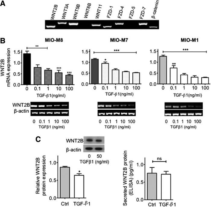
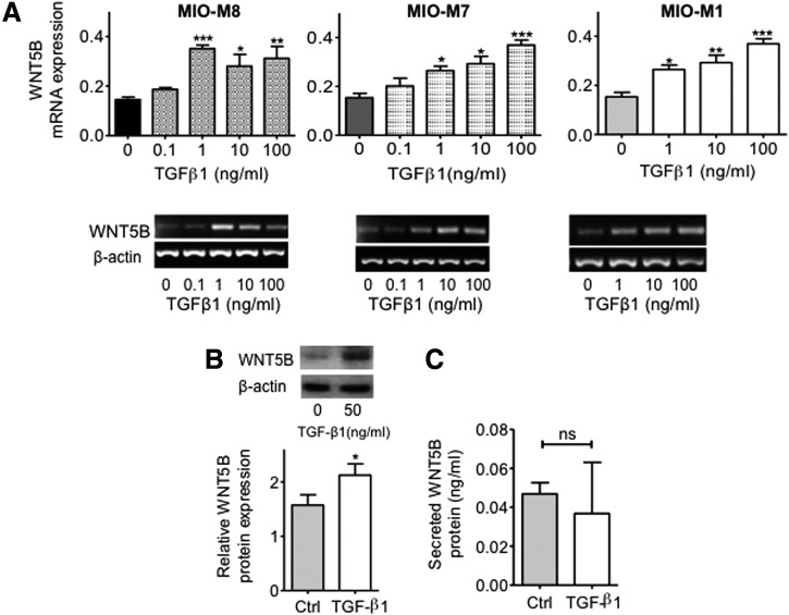
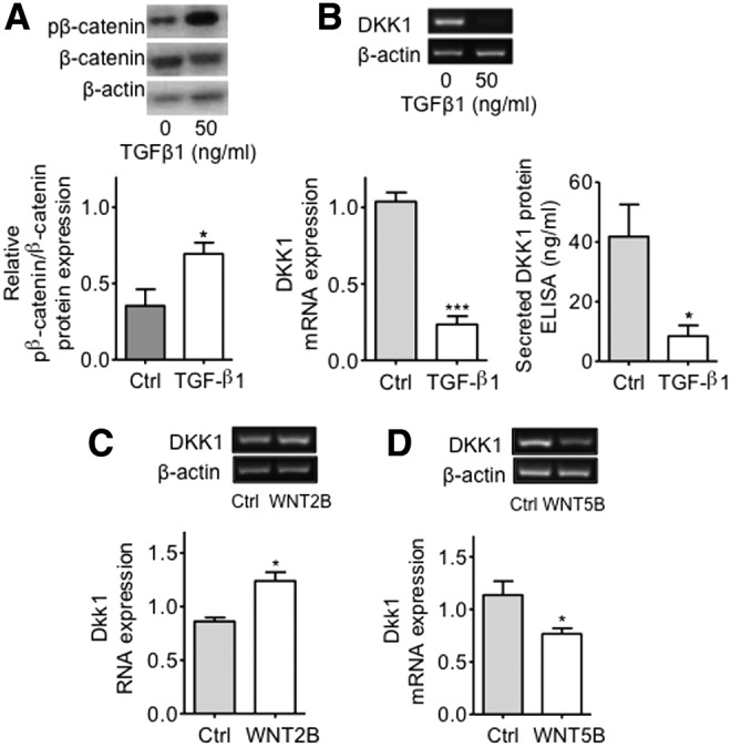
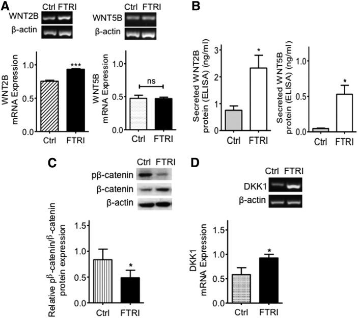
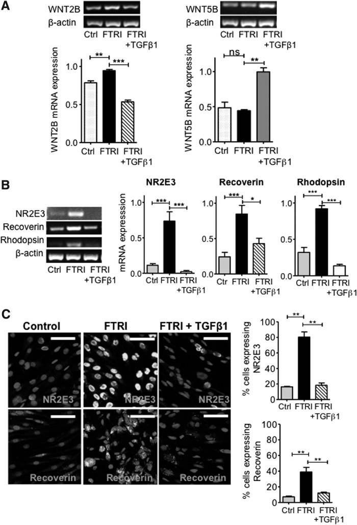
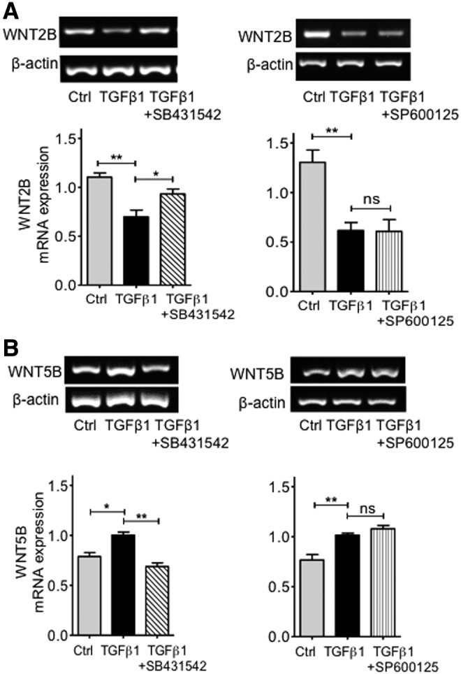
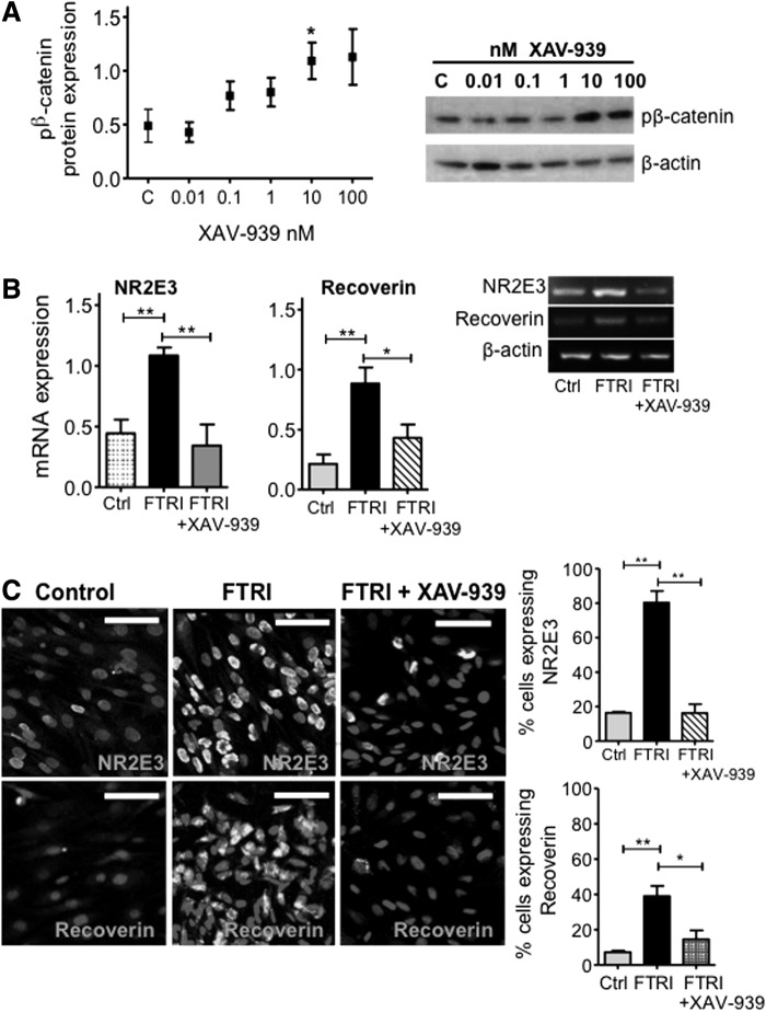
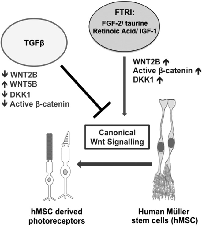
Müller glia are responsible for the retina regeneration observed in zebrafish. Although the human retina harbors Müller glia with stem cell characteristics, there is no evidence that they regenerate the retina after disease or injury. Transforming growth factor-β (TGFβ) and Wnt signaling regulate retinal neurogenesis and inflammation, but their roles in the neural differentiation of human Müller stem cells (hMSC) are not known. We examined hMSC lines in vitro for the expression of various Wnt signaling components and for their modulation by TGFβ1, as well as the effect of this cytokine on the photoreceptor differentiation of these cells. Culture of hMSC with a combination of factors that induce photoreceptor differentiation of hMSC (FGF2, taurine, retinoic acid, and insulin-like growth factor type1; FTRI), markedly upregulated the expression of components of the canonical Wnt signaling pathway, including WNT2B, DKK1, and active β-CATENIN. Although FTRI did not modify mRNA expression of WNT5B, a component of the noncanonical/planar cell polarity Wnt pathway, it upregulated its secretion. Furthermore, TGFβ1 not only decreased WNT2B expression, but also inhibited FTRI-induced photoreceptor differentiation of hMSC, as determined by expression of the photoreceptor markers NR2E3, RHODOPSIN, and RECOVERIN. Inhibition of TGFβ1 signaling by an ALK5 inhibitor prevented TGFβ1-induced changes in the expression of the two Wnt ligands examined. More importantly, inhibition of the canonical WNT signaling by XAV-939 prevented FTRI-induced photoreceptor differentiation. These observations suggest that TGFβ may play a key role in preventing neural differentiation of hMSC and may constitute a potential target for induction of endogenous regeneration of the human retina.


Figures

<b>FIG. 1.</b>
FIG. 1.
Expression of mRNA coding for molecules of the Wnt signaling pathway in human Müller stem cells (hMSC) and modulation of WNT2B expression by transforming growth factor-β (TGFβ1). (A) hMSC express mRNA coding for various components of the canonical and noncanonical Wnt signaling pathway. (B) TGFβ1 downregulation of the expression of mRNA coding for WNT2B occurred in a dose–response manner in three different hMSC lines (MIO-M8, MIO-M7, and MIO-M1) after 7 days culture with concentrations of this cytokine ranging between 0.1 and 100 ng/mL. Histograms represent the mean ± standard error of the mean (SEM) from UV spectrophotometer readings of gel bands. Representative bands are shown below the histograms; n = 3–4. ANOVA test, *P < 0.05; **P < 0.01; ***P < 0.001. (C) A significant decrease in the expression of WNT2B protein was observed by western blot analysis of lysates from cells cultured with 50 ng/mL of TGFβ1. Histograms represent the mean ± SEM of the relative optical density readings of gel bands. Representative bands are shown above the histograms; n = 3. Student's t-test; *P < 0.05. Minimally detectable levels of secreted WNT2B examined by Enzyme-Linked Immunosorbent Assay (ELISA) methods were observed in supernatants of cells cultured in the presence or absence of TGFβ1, and no differences between the two conditions were observed; n = 3. Student's t-test; ns, not significant.
<b>FIG. 2.</b>
FIG. 2.
TGFβ1 modulation on the expression of WNT5B in hMSC. (A) TGFβ1-induced upregulation of the expression of mRNA coding for WNT5B occurred in a dose–response manner in three different hMSC lines examined (MIO-M7, MIO-M8, and MIO-M1). Histograms represent the mean ± SEM from UV spectrophotometer readings of gel bands. Representative bands are shown below the histograms; n = 4. ANOVA test, *P < 0.05; **P < 0.01; ***P < 0.001. (B) Western blot analysis of cell lysates from hMSC showed a marked increase of intracellular protein in cells treated with TGFβ1 at a concentration of 50 ng/mL as compared with control cells. Histograms represent the mean ± SEM of the relative optical density readings of gel bands. Representative bands are shown above the histograms; n = 4. Student's t-test, *P < 0.05. (C) Minimally detectable levels of secreted WNT5B, as examined by ELISA, were observed in supernatants of cells cultured in the presence or absence of TGFβ1, and no differences between the two conditions were observed; n = 3. ns, not significant.
<b>FIG. 3.</b>
FIG. 3.
Modulation of pβ-catenin and DKK1 protein expressions by TGFβ1 and effect of exogenous WNT2B and WNT5B ligands on DKK1 mRNA expression. (A) Western blot analysis revealed that culture of hMSC with 50 ng/mL of TGFβ1 induced a significant upregulation of the ratio of phospho-β-catenin/β-catenin. Histograms represent the mean ± SEM of the relative optical density readings of gel bands. Representative bands are shown above the histograms; n = 5. Student's t-test, *P < 0.05. pβ-catenin=phospho-β-catenin. (B) TGFβ1 caused a significant decrease in DKK1 mRNA expression in hMSC as revealed by RT-PCR analysis. Histograms represent the mean ± SEM from UV spectrophotometer readings of gel bands. Representative bands are shown above the histograms; n = 8. Student's t-test; ***P < 0.001. Secreted DKK1 protein levels as determined by ELISA methods were significantly decreased in culture supernatants of cells treated with 50 ng/mL of TGFβ1 as compared to controls; n = 4. Student's t-test; *P < 0.05. (C) Exogenous addition of recombinant WNT2B into the culture medium induced a significant upregulation of DKK1 mRNA in hMSC; n = 4. Student's t-test, *P < 0.05. (D) Addition of recombinant WNT5B to cells in culture caused a significant downregulation of DKK1 mRNA expression; n = 4. Student's t-test, *P < 0.05. RT-PCR, reverse transcription-polymerase chain reaction.
<b>FIG. 4.</b>
FIG. 4.
Induction of photoreceptor differentiation by FTRI causes changes in the expression of the Wnt signaling components WNT2B, β-catenin, and DKK1 in hMSC. (A) Culture of hMSC with FTRI for 7 days caused a significant increase in the expression of WNT2B mRNA, while no changes in the expression of WNT5B were observed in these cells. Histograms represent the mean ± SEM from UV spectrophotometer readings of gel bands. Representative bands are shown above the histograms; n = 5. Student's t-test, ***P < 0.001. (B) Quantification of the secreted ligands, as measured by ELISA, showed that both WNT2B and WNT5B were significantly increased in culture supernatants of hMSC treated with FTRI for 7 days; n = 3. Student's t-test, *P < 0.05. (C) Western blotting analysis showed that the ratio of phospho-β-catenin/β-catenin was decreased by FTRI treatment of hMSC. Histograms represent the mean ± SEM of the relative optical density readings of gel bands. Representative bands are shown above the histograms; n = 5. Student's t-test, *P < 0.05. pβ-catenin=phospho-β-catenin. (D) A significant increase in the expression of DKK1 mRNA was observed in hMSC cultured with FTRI for 7 days; n = 3. Student's t-test, *P < 0.05. ns, not significant; FTRI, FGF2, taurine, retinoic acid and insulin-like growth factor type1.
<b>FIG. 5.</b>
FIG. 5.
Inhibition of FTRI-induced photoreceptor differentiation of hMSC by TGFβ1. (A) Culture of MIO-M1 cells with FTRI caused an increase in WNT2B mRNA expression, but addition of TGFβ1 to the differentiation medium inhibited this increase; n = 4. ANOVA test, **P < 0.01; ***P < 0.001. FTRI alone did not modify WNT5B mRNA expression, but addition of TGFβ1 to the differentiation cocktail increased WNT5B mRNA expression (similar to that shown above with TGFβ1 alone). Histograms represent the mean ± SEM from UV spectrophotometer readings of gel bands. Representative bands are shown above the histograms; n = 3. ANOVA test, **P < 0.01; ***P < 0.001. (B) Addition of TGFβ1 to hMSC undergoing photoreceptor differentiation with FTRI inhibited the mRNA expression of NR2E3, recoverin, and rhodopsin as compared with FTRI alone; n = 5–8. ANOVA test, *P < 0.05; ***P < 0.001. (C) Immunostaining for NR2E3 and recoverin confirmed that while FTRI alone caused a marked increase in the expression of this photoreceptor protein, addition of TGFβ1 to hMSC cultured with FTRI caused inhibition of photoreceptor differentiation (Alexa 488, fluorescent cells). Cell nuclei counterstained with DAPI (non-fluorescent cell structures). Scale bars 50 μm. Histograms on the right represent the proportion of cells immunostaining for each of the markers following 7-day culture under the different conditions; n = 3. ANOVA test, **P < 0.01.
<b>FIG. 6.</b>
FIG. 6.
Effect of TGFβ1 inhibitors on the expression of the Wnt signaling ligands WNT2B and WNT5B by hMSC. (A) Addition of the TGFβ type I receptor (ALK5) inhibitor SB431542 (10 μM) to cells cultured with TGFβ1 antagonized the inhibitory effects of this cytokine on WNT2B mRNA expression; n = 5. ANOVA test; *P < 0.05; **P < 0.01. In contrast, addition of the JNK inhibitor SP600125 (20 μM) to cells cultured in the presence of TGFβ1 did not modify the effect of this cytokine on WNT2B gene expression. Histograms represent the mean ± SEM from UV spectrophotometer readings of gel bands. Representative bands are shown above the histograms; n = 3. ANOVA test, **P < 0.01. (B) While the ALK5 inhibitor SB431542 antagonized the upregulation of WNT5B mRNA by TGFβ1; n = 3. ANOVA test; *P < 0.05; **P < 0.01, the JNK inhibitor SP600125 did not modify the effects of this cytokine on the expression of this ligand gene; n = 4. ANOVA test, **P < 0.01; ns, not significant.
<b>FIG. 7.</b>
FIG. 7.
Effect of inhibition of β-CATENIN on the photoreceptor differentiation of hMSC. (A) Dose–response increase in pβ-CATENIN expression by hMSC cultured with concentrations of XAV939 ranging between 0.01 and 100 nM. Western blot bands on the right show that XAV-939 significantly upregulated the expression of pβ-CATENIN protein at concentrations of 10 and 100 nM; n = 4. Student's t-test, *P < 0.05. pβ-catenin=phospho-β-catenin. (B) Addition of XAV-939 to hMSC cultured with FTRI inhibited the differentiation of these cells into photoreceptors, as judged by the expression of mRNA coding for NR2E3 and RECOVERIN. Histograms on the left represent the mean ± SEM from UV spectrophotometer readings of gel bands. Representative bands are shown on right of the histograms; n = 5–7. ANOVA test; *P < 0.05; **P < 0.01. (C) Confocal images confirmed that addition of XAV-939 to hMSC cultured in the presence of FTRI caused a decrease in the expressions of NR2E3 and recoverin, which is upregulated by FTRI alone (Alexa 488, fluorescent cells). Cell nuclei counterstained with DAPI (non-fluorescent cell structures). Scale bars 50 μm. Histogram represents the percentage of cells stained with NR2E3 following 7 days culture under the different conditions; n = 3. ANOVA test; *P < 0.05; **P < 0.01.
<b>FIG. 8.</b>
FIG. 8.
TGFβ1 inhibits the canonical Wnt signaling pathway necessary for the photoreceptor differentiation of hMSC in vitro. Schematic illustration summarizing the interactions of FTRI, WNT2B, WNT5B, TGFβ1, and DKK1 in hMSC. FTRI, which induces photoreceptor differentiation of hMSC, activated the canonical Wnt signaling pathway in these cells. Addition of TGFβ1 to cells cultured with FTRI caused inhibition of the canonical Wnt signaling and consequently inhibited the photoreceptor differentiation of hMSC in vitro.

References

    1. Bernardos RL, Barthel LK, Meyers JR. and Raymond PA. (2007). Late-stage neuronal progenitors in the retina are radial Müller glia that function as retinal stem cells. J Neurosci 27:7028–7040 - PMC - PubMed
    1. Lawrence JM, Singhal S, Bhatia B, Keegan DJ, Reh TA, Luthert PJ, Khaw PT. and Limb GA. (2007). MIO-M1 cells and similar Müller glial cell lines derived from adult human retina exhibit neural stem cell characteristics. Stem Cells 25:2033–2043 - PubMed
    1. Jayaram H, Jones MF, Eastlake K, Cottrill PB, Becker S, Wiseman J, Khaw PT. and Limb GA. (2014). Transplantation of photoreceptors derived from human Müller glia restore rod function in the P23H rat. Stem Cells Transl Med 3:323–333 - PMC - PubMed
    1. Singhal S, Bhatia B, Jayaram H, Becker S, Jones MF, Cottrill PB, Khaw PT, Salt TE. and Limb GA. (2012). Human Müller glia with stem cell characteristics differentiate into retinal ganglion cell (RGC) precursors in vitro and partially restore RGC function in vivo following transplantation. Stem Cells Transl Med 1:188–199 - PMC - PubMed
    1. Becker S, Singhal S, Jones MF, Eastlake K, Cottrill PB, Jayaram H. and Limb GA. (2013). Acquisition of RGC phenotype in human Müller glia with stem cell characteristics is accompanied by upregulation of functional nicotinic acetylcholine receptors. Mol Vis 19:1925–1936 - PMC - PubMed

MeSH terms

LinkOut - more resources