

Scaffold-free, Human Mesenchymal Stem Cell-Based Tissue Engineered Blood Vessels
- PMID: 26456074
- PMCID: PMC4600980
- DOI: 10.1038/srep15116
Scaffold-free, Human Mesenchymal Stem Cell-Based Tissue Engineered Blood Vessels
Abstract
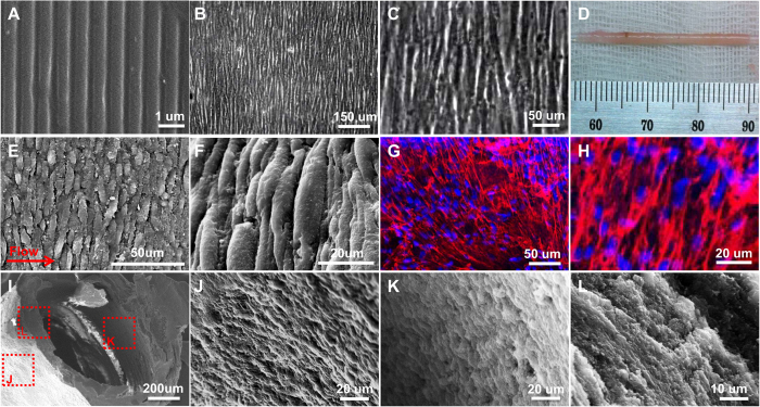
Tissue-engineered blood vessels (TEBV) can serve as vascular grafts and may also play an important role in the development of organs-on-a-chip. Most TEBV construction involves scaffolding with biomaterials such as collagen gel or electrospun fibrous mesh. Hypothesizing that a scaffold-free TEBV may be advantageous, we constructed a tubular structure (1 mm i.d.) from aligned human mesenchymal cell sheets (hMSC) as the wall and human endothelial progenitor cell (hEPC) coating as the lumen. The burst pressure of the scaffold-free TEBV was above 200 mmHg after three weeks of sequential culture in a rotating wall bioreactor and perfusion at 6.8 dynes/cm(2). The interwoven organization of the cell layers and extensive extracellular matrix (ECM) formation of the hMSC-based TEBV resembled that of native blood vessels. The TEBV exhibited flow-mediated vasodilation, vasoconstriction after exposure to 1 μM phenylephrine and released nitric oxide in a manner similar to that of porcine femoral vein. HL-60 cells attached to the TEBV lumen after TNF-α activation to suggest a functional endothelium. This study demonstrates the potential of a hEPC endothelialized hMSC-based TEBV for drug screening.
Figures


References
-
- Benam K. H. et al. Engineered in vitro disease models. Annual Review of Pathology: Mechanisms of Disease 10, 195–262 (2015). - PubMed
-
- Esch M. B. K. T. & Shuler M. L. The role of body-on-a-chip devices in drug and toxicity studies. 15, 55–72 (2011). - PubMed
-
- “R & D costs are on the rise. (News). (new drug development) (Brief Article).” Medical Marketing & Media. Haymarket Media. 2003. HighBeam Research. Date of access: 14/03/2015 < http://www.highbeam.com/doc/1G1-102908512.html>.
-
- Bhadriraju K. & Chen C. S. Engineering cellular microenvironments to improve cell-based drug testing. Drug Discovery Today 11, 612–620 (2002). - PubMed
-
- Pampaloni F., Reynaud E. G. & Stelzer E. H. K. The third dimension bridges the gap between cell culture and live tissue. Nat Rev Mol Cell Biol 8, 839–845 (2007). - PubMed
Publication types
MeSH terms
Substances
Grants and funding
LinkOut - more resources
Full Text Sources
Other Literature Sources
