. 2015 Dec;171(5):798-812.
doi: 10.1111/bjh.13780. Epub 2015 Oct 12.

Synergistic anti-myeloma activity of the proteasome inhibitor marizomib and the IMiD immunomodulatory drug pomalidomide

Affiliations

Synergistic anti-myeloma activity of the proteasome inhibitor marizomib and the IMiD immunomodulatory drug pomalidomide

Deepika S Das et al. Br J Haematol. 2015 Dec.

Abstract

The proteasome inhibitor bortezomib is an effective therapy for the treatment of relapsed and refractory multiple myeloma (RRMM); however, prolonged treatment can be associated with toxicity, peripheral neuropathy and drug resistance. Our earlier studies showed that the novel proteasome inhibitor marizomib is distinct from bortezomib in its chemical structure, mechanisms of action and effects on proteasomal activities, and that it can overcome bortezomib resistance. Pomalidomide, like lenalidomide, has potent immunomodulatory activity and has been approved by the US Food and Drug Administration for the treatment of RRMM. Here, we demonstrate that combining low concentrations of marizomib with pomalidomide induces synergistic anti-MM activity. Marizomib plus pomalidomide-induced apoptosis is associated with: (i) activation of caspase-8, caspase-9, caspase-3 and PARP cleavage, (ii) downregulation of cereblon (CRBN), IRF4, MYC and MCL1, and (iii) suppression of chymotrypsin-like, caspase-like, and trypsin-like proteasome activities. CRBN-siRNA attenuates marizomib plus pomalidomide-induced MM cells death. Furthermore, marizomib plus pomalidomide inhibits the migration of MM cells and tumour-associated angiogenesis, as well as overcomes cytoprotective effects of bone marrow microenvironment. In human MM xenograft model studies, the combination of marizomib and pomalidomide is well tolerated, inhibits tumour growth and prolongs survival. These preclinical studies provide the rationale for on-going clinical trials of combined marizomib and pomalidomide to improve outcome in patients with RRMM.

Keywords: cancer and drug therapy; marizomib; multiple myeloma; pomalidomide.


Conflict of interest statement

Conflict-of-interest disclosure MT is an employee of Triphase Accelerator; KCA is on Advisory board of Celgene, Onyx, Gilead, Sonofi Aventis and is a Scientific founder of Acetylon and Oncopep; DC is consultant to Triphase Accelerator. The remaining authors declare no competing financial interests.

Figures

Figure 1
Figure 1. Combination of low doses of marizomib and pomalidomide induces synergistic cell death in MM cell lines, patient MM cells but not normal PBMCs
(A–D) MM-cell lines (MM.1S, ANBL6.BR, ARP-1, and MM.1R) were pretreated with or without pomalidomide (POM) for 24 h; marizomib was then added for an additional 24 h, followed by assessment for cell viability using the MTT assay. The experiments with single agents and respective combinations were carried out simultaneously. Isobologram analysis shows the synergistic cytotoxic effect of marizomib and pomalidomide. The graph (right panel) is derived from the values given in the table (left panel). A combination index (CI) <1 indicates synergy. (E) Purified patient MM cells (CD138-positive) were pretreated with pomalidomide for 24 h; marizomib was then added for an additional 24 h, followed by cell death analysis using CellTiter-Glo assay. Data are mean ± SE of triplicate samples (p < 0.05 for all patient samples, for single agent vs combination treated samples). (F) Peripheral blood mononuclear cells (PBMCs) from healthy donors were treated (as in panel A) with indicated concentrations of marizomib, pomalidomide or marizomib plus pomalidomide, and then analysed for viability using the WST-1 assay. Data are mean ± SE (n=5). Fa = fraction of cells affected.
Figure 2
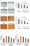
Figure 2. Combined low doses of marizomib and pomalidomide block migration, tubule formation, and cytoprotective effects of BMSCs and pDCs
(A) Migration assay: MM.1S cells were pretreated with pomalidomide for 12h, and then marizomib was added for an additional 6h; cells were more than 90% viable at this time point. The cells were washed and cultured in serum-free medium. After 2h incubation, cells were plated on a fibronectin-coated polycarbonate membrane in the upper chamber of transwell inserts and exposed for 4h to serum-containing medium in the lower chamber. Cells migrating to the bottom face of the membrane were fixed with 90% ethanol and stained with crystal violet. A total of 3 randomly selected fields were examined for cells that had migrated from the top to the bottom chambers. (Left panel) Image is representative of 2 experiments with similar results. (Right panel) The bar graph represents quantification of migrated cells. Data are mean ± SE (p < 0.05). (B) Human vascular endothelial cells were cultured in the presence or absence of combined low doses of marizomib plus pomalidomide for 48 h, and then assessed for in vitro angiogenesis using matrigel capillary-like tube structure formation assays (Left panel). Image is representative from 3 experiments with similar results. In vitro angiogenesis is reflected by capillary tube branch formation (dark brown). (Right panel) The bar graph represents quantification of capillary-like tube structure formation in response to indicated agents: Branch points in several random view fields/well were counted, values were averaged, and statistically significant differences were measured using Student’s t test. (C–D) MM.1S cells were cultured in bone marrow stromal cells (BMSC)- or plasmacytoid dendritic cells (pDC)-coated or uncoated wells with control medium, marizomib, pomalidomide or marizomib plus pomalidomide. Cell proliferation was assessed by Brdu colorimetric assay. Data are mean ± SE (n=3; p < 0.05, for control vs combination treated samples).
Figure 3
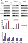
Figure 3. Marizomib plus pomalidomide-induced apoptosis in MM cells is associated with activation of caspases
(A) MM.1S, RPMI-8226 or Dox-40 cells were pre-treated with or without pomalidomide for 24 h, and then marizomib was added for an additional 24 h; protein lysates were subjected to immunoblot analysis with anti-PARP, anti-caspase-3, anti-caspase-8, anti-caspase-9 or anti-GAPDH FL, full length; CF, cleaved fragment. Blots shown are representative of 3 independent experiments. (B) MM.1S cells were pre-treated with or without pomalidomide for 24 h, and then marizomib was added for an additional 24 h; protein lysates were subjected to measurement of caspase-8 or caspase-9 enzymatic activity using fluorometric kit. Data are mean ± SE (n=3; p < 0.05, for single agent vs combination treated samples) (C) MM.1S cells were treated with indicated agents (as in panel ‘A’) in the presence or absence of biochemical inhibitors of caspase-3, caspase-8, caspase-9, or pan-caspase and then analyzed for apoptosis using WST-1 assay. Data are mean ± SE (n=3; p < 0.05, for control vs combination treated samples).
Figure 4
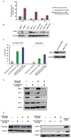
Figure 4. Mechanisms mediating anti-MM activity of marizomib plus pomalidomide
(A) MM.1S cells were pretreated with or without pomalidomide for 12 h, and then marizomib was added for an additional 12 h; cells were harvested and cytosolic extracts were then analysed for chymotrypsin- like (CT-L), caspase-like (C-L) and trypsin-like (T-L) proteasome activities. Results are represented as percentage inhibition in proteasome activities in drug-treated versus vehicle control. Data are mean ± SE (n=3; p < 0.05, for single agent vs combination treated samples) (B) Total protein lysates from the indicated MM cell lines and normal healthy donor PBMCs were subjected to immunoblot analysis with anti-CRBN or anti-GAPDH Abs. (C) MM.1S cells were transfected with siRNA-CRBN or scr-siRNA for 24h. Transfected cells were pretreated with or without pomalidomide for 24h; marizomib was then added for an additional 24h, followed by analysis for apoptosis using WST-1 assay. Data are mean ± SE (n=3; p < 0.05, for single agent vs combination treated samples) Immunoblot shows CRBN expression in cells transfected with scr-siRNA or CRBN-siRNA. (D and E) MM.1S cells were treated with indicated agents (as treated in panel ‘A’); protein lysates were subjected to immunoblot analysis with anti-CRBN, anti-IRF4, anti-MYC, anti-MCL1, anti-HSP90AA1, anti-HSPA1A, anti-HSPB1 or anti-GAPDH Abs.
Figure 5
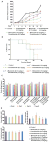
Figure 5. Combination of low doses of marizomib and pomalidomide inhibits human plasmacytoma growth in CB-17 SCID mice
(A) Mice bearing human MM.1S MM tumours (n=5/group) were treated with vehicle, marizomib (0.15 mg/kg; orally), pomalidomide (0.5 mg/kg or 2.5 mg/kg; orally), or marizomib plus pomalidomide (orally) at the indicated doses for 24 days on a twice-weekly schedule for marizomib and for 4 consecutive days weekly for pomalidomide. Bars represent mean ± SE. (B) Kaplan-Meier plots showing survival for mice treated with marizomib, pomalidomide, or marizomib plus pomalidomide at the indicated doses. Marizomib plus pomalidomide-treated mice show significantly increased survival (p < 0.05, Logrank test for trend) compared with the untreated group. (C) Vehicle-treated control mice, as well as mice in the marizomib, pomalidomide, or marizomib plus pomalidomide-treated cohorts, were weighed every week. The average changes in body weight are shown. (D) Mice were treated with vehicle, marizomib, pomalidomide, or marizomib plus pomalidomide (as in panel ‘A’) for 24 days; blood samples were then obtained and subjected to analysis for serum bilirubin, hemoglobin, and creatinine levels using Quantichrom Creatinine, Bilirubin, and Haemoglobin Assay kit (BioAssay Systems, Hayward, CA, USA).
Figure 6
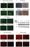
Figure 6. Effect of marizomib plus pomalidomide on apoptosis, MCL1, IRF4 and neovascularization in vivo in xenografted MM tumours
(A and B) Apoptotic cells in sections of tumours harvested from vehicle control-, marizomib-, pomalidomide-, or marizomib (0.15 mg/kg) plus pomalidomide (2.5 mg/kg)-treated mice were identified by immunostaining for activated caspase-3 (green cells) or TUNEL. Tumour sections were obtained on day 24. Images were obtained with a Leica SP5X laser scanning confocal microscope (40x magnification). (C) Tumours harvested from mice (as in panel ‘A’) were immunostained with Ki67 Abs (40x magnification). (D) Tumour lysates from control and drug-treated mice were subjected to immunoblot analysis using anti-MCL1, anti-IRF4, or anti-GAPDH Abs. Lanes 1 to 6 represent lysates of tumours harvested from mice receiving the following treatments: lane 1, vehicle alone (control); lane 2, marizomib (0.15 mg/kg); lane 3, pomalidomide (0.5 mg/kg); lane 4, pomalidomide (2.5 mg/kg); lane 5, marizomib (0.15 mg/kg) plus pomalidomide (2.5 mg/kg); and lane 6, marizomib (0.15 mg/kg) plus pomalidomide (0.5mg/kg). (E and F) Tumours harvested from mice (as in panel ‘A’) were immunostained with Factor VIII or VEGFR1 Abs. Images were obtained with a Leica SP5X laser scanning confocal microscope (40x magnification). Photographs (A, B, C, E, and F) shown are representative of similar observations in 2 mice receiving the same treatment.

Similar articles

Cited by

References

    1. Anderson KC. Targeted therapy of multiple myeloma based upon tumor-microenvironmental interactions. Exp Hematol. 2007;35:155–162. - PubMed
    1. Anderson KC. The 39th David A. Karnofsky Lecture: bench-to-bedside translation of targeted therapies in multiple myeloma. J Clin Oncol. 2012;30:445–452. - PMC - PubMed
    1. Arendt C, Hochstrasser M. Identification of the yeast 20S proteasome catalytic centers and subunit interactions required for active-site formation. Proc Natl Acad Sci U S A. 1997;94:7156–7161. - PMC - PubMed
    1. Atrash S, Tullos A, Panozzo S, Bhutani M, Van Rhee F, Barlogie B, Usmani SZ. Cardiac complications in relapsed and refractory multiple myeloma patients treated with carfilzomib. Blood Cancer J. 2015;5:e272. - PMC - PubMed
    1. Avet-Loiseau H, Attal M, Moreau P, Charbonnel C, Garban F, Hulin C, Leyvraz S, Michallet M, Yakoub-Agha I, Garderet L, Marit G, Michaux L, Voillat L, Renaud M, Grosbois B, Guillerm G, Benboubker L, Monconduit M, Thieblemont C, Casassus P, Caillot D, Stoppa AM, Sotto JJ, Wetterwald M, Dumontet C, Fuzibet JG, Azais I, Dorvaux V, Zandecki M, Bataille R, Minvielle S, Harousseau JL, Facon T, Mathiot C. Genetic abnormalities and survival in multiple myeloma: the experience of the Intergroupe Francophone du Myelome. Blood. 2007;109:3489–3495. - PubMed

Publication types

MeSH terms