. 2015 Nov 3;22(5):907-21.
doi: 10.1016/j.cmet.2015.09.009. Epub 2015 Oct 8.

Identification of AMPK Phosphorylation Sites Reveals a Network of Proteins Involved in Cell Invasion and Facilitates Large-Scale Substrate Prediction

Affiliations

Identification of AMPK Phosphorylation Sites Reveals a Network of Proteins Involved in Cell Invasion and Facilitates Large-Scale Substrate Prediction

Bethany E Schaffer et al. Cell Metab. .

Abstract

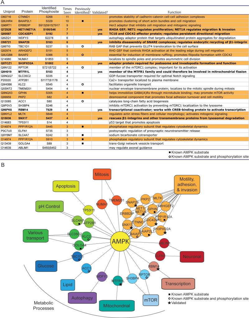
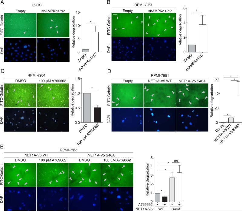
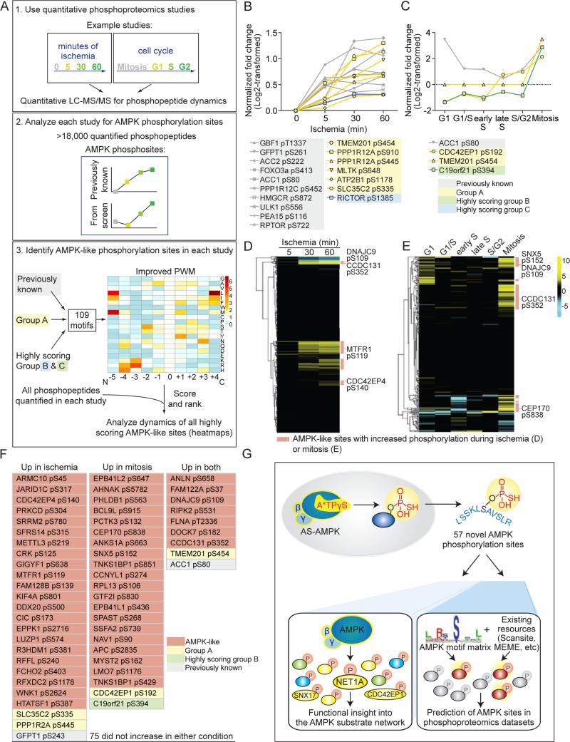
AMP-activated protein kinase (AMPK) is a central energy gauge that regulates metabolism and has been increasingly involved in non-metabolic processes and diseases. However, AMPK's direct substrates in non-metabolic contexts are largely unknown. To better understand the AMPK network, we use a chemical genetics screen coupled to a peptide capture approach in whole cells, resulting in identification of direct AMPK phosphorylation sites. Interestingly, the high-confidence AMPK substrates contain many proteins involved in cell motility, adhesion, and invasion. AMPK phosphorylation of the RHOA guanine nucleotide exchange factor NET1A inhibits extracellular matrix degradation, an early step in cell invasion. The identification of direct AMPK phosphorylation sites also facilitates large-scale prediction of AMPK substrates. We provide an AMPK motif matrix and a pipeline to predict additional AMPK substrates from quantitative phosphoproteomics datasets. As AMPK is emerging as a critical node in aging and pathological processes, our study identifies potential targets for therapeutic strategies.


Figures

Figure 1
Figure 1. Screening strategy to identify AMPKα1 andα2 substrates and phosphorylation sites in cells
(A) Schematic of the peptide-capture technique used to identify analog-specific (AS) AMPKα1 and α2 substrates and phosphorylation sites in whole cells. AS-AMPK uses A*TPγS, a bulky ATP analog, to thiophosphorylate substrates. Upper panel: thiophosphorylated substrates are alkylated by p-nitrobenzyl mesylate (PNBM) and recognized by an antibody to the thiophosphate moiety (thioP). Lower panel: thiophosphorylated peptides are captured on a resin, eluted, and identified using liquid chromatography-tandem mass spectrometry (LC-MS/MS). “2DG”, 2-deoxy-D-glucose. (B) HA-tagged AS-AMPKα1 and α2 thiophosphorylate endogenous substrates in U2OS cells without over-expression of the β and γ subunits. Cells were serum-starved for 2 hours and stimulated for 5 minutes with 100 mM 2DG, then incubated with A*TPγS. Whole cell lysates were analyzed for the presence of thiophosphorylation (thioP) and exogenous AMPK subunits (HA tag). (C) HA-tagged AS-AMPKα1 and α2 thiophosphorylate endogenous substrates under different AMPK-activating conditions. Whole cell lysates were analyzed for the presence of thiophosphorylation (thioP) and AMPKα (HA tag, AMPKα1, AMPKα2). First panel: 2 hours of serum-starvation with 5 minutes of 100 mM 2DG; second panel: 15 minutes of 50 mM 2DG; third panel: 30 minutes of 300 μM A769662. Representative of 6, 1, and 3 independent experiments for 2DG (−) serum, 2DG, and A769662, respectively. “Empty”, Empty vector; “α1WT”, WT-AMPKα1; “α1AS”, AS-AMPKα1, “α2WT”, WT-AMPKα2, “α2AS”, AS-AMPKα2. (D) Summary of mass spectrometry datasets. AMPK-activating conditions as in Figure 1C. See Figure S1G and Supplementary List 1 for more information. “Empty”, Empty vector. (E) Known AMPK substrates identified in multiple AS-AMPK datasets. Underlined and bold residues, phosphorylated sites on the identified phosphopeptide (more than one is shown if the phosphopeptide had multiple or ambiguous phosphorylation site identification). “Phosphosite” column, identified phosphorylation site corresponding to the known AMPK site. “|”, ambiguous site identification. S722, known AMPK site on RAPTOR; S237, known site on TBC1D1; S366, known site on BAIAP2.
Figure 2
Figure 2. Identification of high confidence AS-AMPK substrates with a tailored pipeline
(A) Schematic of the pipeline used to identify AS-AMPK substrates in LC-MS/MS datasets. See Supplemental Experimental Procedures. Phosphopeptides only found in ASAMPK datasets were classified as Group A, B, or C based on the number of biological samples in which they were identified. “α1WT”, “α2WT”, “α1AS”, “α2AS”, WT or ASAMPKα1 or α2. (B-D) Logo motif of the most common phosphorylation sites on each phosphopeptide from Group A (B), Group B (C) and Group C (D). The established in vitro AMPK phosphorylation motif displayed below Group A is modified with permission from (Gwinn et al., 2008), and was generated in that study using a positional scanning peptide library. Green, hydrophobic residues; red, basic; yellow, acidic; blue, neutral polar. See Figure S2A and Supplemental Experimental Procedures for selection of the most common phosphorylation sites.
Figure 3
Figure 3. AS-AMPK directly phosphorylates several high confidence substrates, including SNX17 and NET1A
(A-D) AS-AMPK thiophosphorylates SNX17, NET1A, CDC42EP1, and SH3PXD2A. Tagged proteins were over-expressed in empty vector or AMPKα-expressing (HA-tagged WT or AS-AMPKα1 or α2) U2OS cell lines, immunoprecipitated, and analyzed by western blot for the presence of thiophosphorylation. AMPK was activated in all conditions with 15 minutes of 50 mM 2DG. Representative of 3, 1, 1, and 2 independent experiments, respectively. (E) Mass spectrometry-predicted AMPK phosphorylation sites and corresponding phosphopeptides for SNX17, NET1A, and CDC42EP1. Labeled as in Figure 1E. S100 on NET1 (S46 on the short isoform NET1A) was used as the NET1/NET1A site as its surrounding motif resembled the AMPK motif better than that of T99. (F-H) AS-AMPK thiophosphorylates SNX17, NET1A, and CDC42EP1 at the identified residues. Tagged WT and predicted phosphorylation site mutants of the indicated substrates were over-expressed in U2OS AS-AMPKα2 and α1 cell lines, immunoprecipitated, and analyzed as in Figures 3A-D. The phosphorylation motif for the predicted residue is shown. The phosphorylated residue is underlined and bold. Color-coding as in Figure 2B. Each panel representative of 2 independent experiments. (I) AMPK phosphorylates S437 on SNX17 endogenously. AMPK was activated in U2OS cells stably expressing an shRNA against AMPKα1 and α2 or empty vector control. Phosphorylation of the known substrates ACC1 S80 and PPP1R12C S452 are shown as controls for AMPK activation. Note that there is still some degree of AMPK substrate phosphorylation in cells with stable knockdown of AMPKα1 and α2, probably due to residual AMPK expression in these cells. “-“, no drug (DMSO vehicle control); A, A769662, 300 μM for 30 minutes. Representative of 2 independent experiments. (J) Specific activation of AMPK decreases SNX17 protein levels. AMPK was activated in U2OS cells with 300 μM of A769662 for the indicated amount of time. Representative of 2 independent experiments. (K) Over-expressed NET1A is phosphorylated at S46 in response to endogenous AMPK activation. NET1A-V5 WT or S46A was expressed in a doxycycline-inducible manner in U2OS cell lines. NET1A-V5 WT was also expressed in U2OS cell lines with shRNA-knockdown of both AMPKα1 and α2. Cells were serum-starved overnight, which was important to decrease basal NET1A phosphorylation, and NET1A-V5 expression was induced by 2 hours of doxycycline exposure (see Supplemental Experimental Procedures). AMPK was activated with 300 μM A769662 for 30 minutes. Following NET1A-V5 immunoprecipitation, samples were immunoblotted with an AMPK substrate motif antibody. Representative of 3 independent experiments.
Figure 4
Figure 4. Many high confidence AMPK substrates have known roles in cell motility, adhesion, and invasion
(A) All Group A substrates and the most frequently identified phosphorylation site(s) on the phosphopeptide. “|”, ambiguity in the mass spectrometry placement of the phosphorylation site; “Times seen”, number of biological samples the phosphopeptide was identified in; “Previously identified” indicates whether the phosphorylation site (open circle) or the protein (but not phosphorylation site) (closed square) was a previously known AMPK substrate; “Validated?” indicates whether the protein was validated as a substrate of AS-AMPK (Figures 3 and S3). Orange background, proteins with known roles in cell motility, adhesion, or invasion (see Figure S4B); note that 2 different sites were identified on PPP1R12A. Bold type, validated substrates. (B) Many Group A substrates are proteins involved in cell motility, adhesion, and invasion. Twelve substrates involved in motility, adhesion and invasion were classified by mining the literature (Figure S4B), while an additional 2 were identified using a curated list of GO Terms (Figures S4A and S4B).
Figure 5
Figure 5. AMPK phosphorylation of NET1A inhibits extracellular matrix (ECM) degradation
(A) Knockdown of AMPK increases ECM degradation in U2OS cells. Cells stably expressing an shRNA against both AMPKα1 and α2 or empty vector control (Empty) cells were cultured on Fluorescein isothyocyanite (FITC)-conjugated gelatin-coated coverslips for 3 days. Fixed cells were stained for DAPI and analyzed. Grey arrowheads indicate points of gelatin degradation. Within each experiment, approximately 15-20 20× fields per sample were quantified and averaged; displayed images are 40x. Error bars represent mean +/− SEM of the averaged values from 6 independent experiments; the control samples in 4 of the experiments were the same used in 4 of the experiments in Figure S5G. *p < 0.05 by two-tailed Wilcoxon matched-pairs signed ranked test; scale bar = 50 μM. (B) Knockdown of AMPK increases ECM degradation in RPMI-7951 cells. Cells stably expressing an shRNA against both AMPKα1 and α2 or empty vector control cells were analyzed and results are represented as in Figure 5A. Seven independent experiments were quantified. (C) Activation of AMPK inhibits ECM degradation. RPMI-7951 cells were plated on FITC-conjugated gelatin-coated coverslips for 3-4 hours prior to administration of 100 μM A769662 or DMSO vehicle control for 16 hours. Analysis and results are represented as in Figure 5A. Six independent experiments were quantified. (D) Loss of the AMPK phosphorylation site on NET1A increases ECM degradation. RPMI-7951 cells expressing similar levels of doxycycline-inducible NET1A-V5 WT or S46A (Figures S5C and S5D) were plated on FITC-conjugated gelatin coated coverslips and allowed to adhere overnight. 2 μg/ml doxycycline was added, and cells were cultured for an additional 2 days. Analysis and results are as in Figure 5A. Six independent experiments were quantified. (E) Activation of AMPK inhibits ECM degradation in the presence of WT, but not S46A, NET1A. RPMI-7951 cells expressing similar levels of doxycycline-inducible NET1A-V5 WT or S46A (Figures S5C and S5D) were plated on FITC-conjugated gelatin coated coverslips for 3 hours prior to addition of 2 μg/ml doxycycline and either 100 μM A769662 or DMSO vehicle control for 20 hours. Media and drugs were replaced with fresh stocks after 10 hours. Analysis and results are represented as in Figure 5A. Six independent experiments were quantified; ns, not significant.
Figure 6
Figure 6. Use of the AMPK phosphorylation motif to rank phosphorylation sites identified at low frequency in the screen
(A) Schematic of the pipeline used to score resemblance to the AMPK phosphorylation motif. The logo motif (1.) represents the phosphorylation motif of 50 known in vivo AMPK substrates. See Figure 2B for amino acid color-coding. The heat map (2. and 5.) represents the standardized frequencies of the 50 known AMPK substrates, with red indicating enrichment and blue indicating depletion in the AMPK motif compared with background (see Figure S6A). (B) Ranked list of the scored motifs corresponding to the Group A (yellow lines), B (green lines), and C (blue lines) phosphorylation sites. The logo motif for each quartile of ranked motifs is shown. Scores of interest are noted. (C) Nine highly scoring Group B and C phosphorylation sites are on proteins with known roles in cell motility, adhesion, and invasion. When combined with Group A, this totals 24 phosphorylation sites on 22 proteins involved in these processes (2 sites are on PPP1R12C and ERBB2IP).
Figure 7
Figure 7. In silico analysis of AMPK network dynamics and prediction of AMPK phosphorylation sites
(A) 1. General schematic of quantitative phosphoproteomic studies. 2. Datasets were queried for the presence of known AMPK phosphorylation sites and their dynamics during the biological processes analyzed. 3. A PWM constructed from 109 AMPK phosphorylation sites from both the literature and this study (Supplemental List 6) was generated to score and rank the motifs surrounding each quantified site in 1. The dynamics of sites with motifs scoring above the cutoff (Figure S7F) were analyzed. (B) Seventeen AMPK phosphorylation sites present in the ischemia study (Mertins et al., 2014) increased significantly during ischemia (significance determined in (Mertins et al., 2014)). Sites in yellow are from Group A; blue, highly scoring Group C substrates; grey, previously known. Phosphorylation site location was standardized to isoform 1 in Uniprot. The 5, 30, and 60 minute time-points were standardized to the 0 minute time-point and log2-transformed in (Mertins et al., 2014). (C) Four AMPK phosphorylation sites present in the cell cycle dataset (Olsen et al., 2010) increased at least 2-fold (following log2-transformation) during mitosis. Sites with yellow lines are from Group A; green, highly scoring Group B substrates; grey, previously known. Site location was standardized to isoform 1 in Uniprot. All timepoints were standardized to an asynchronously cycling population, normalized to protein level, and log2-transformed in (Olsen et al., 2010). (D) 630 AMPK-like phosphorylation sites were present in the ischemia dataset, and a subset are dynamically phosphorylated (Supplemental List 7). Yellow, relative increase; blue, relative decrease compared to the 0 minute time-point. The time-points were standardized as in Figure 7B and (Mertins et al., 2014). (E) 266 AMPK-like phosphorylation sites were present in the cell cycle dataset, and a subset are dynamically phosphorylated (Supplemental List 7). Yellow, relative increase; blue, relative decrease compared to an asynchronously cycling population. The relative changes were standardized as in Figure 7C and (Olsen et al., 2010). (F) Highly scoring AMPK-like sites quantified in both the ischemia and cell cycle datasets and whose phosphorylation increased during ischemia and/or mitosis. A total of 131 quantified AMPK-like sites were present in both datasets (Supplemental List 7). Phosphorylation sites are as reported in the ischemia study (based on Protein GI Accessions). (G) Summary of the proteomic and in silico approaches used here to identify AMPK phosphorylation sites and understand the AMPK functional network.

References

    1. Amato S, Liu X, Zheng B, Cantley L, Rakic P, Man HY. AMP-activated protein kinase regulates neuronal polarization by interfering with PI 3-kinase localization. Science. 2011;332:247–251. - PMC - PubMed
    1. Andersson U, Filipsson K, Abbott CR, Woods A, Smith K, Bloom SR, Carling D, Small CJ. AMP-activated protein kinase plays a role in the control of food intake. J Biol Chem. 2004;279:12005–12008. - PubMed
    1. Apfeld J, O'Connor G, McDonagh T, DiStefano PS, Curtis R. The AMP-activated protein kinase AAK-2 links energy levels and insulin-like signals to lifespan in C. elegans. Genes Dev. 2004;18:3004–3009. - PMC - PubMed
    1. Bailey TL, Boden M, Buske FA, Frith M, Grant CE, Clementi L, Ren J, Li WW, Noble WS. MEME SUITE: tools for motif discovery and searching. Nucleic Acids Res. 2009;37:W202–208. - PMC - PubMed
    1. Banko MR, Allen JJ, Schaffer BE, Wilker EW, Tsou P, White JL, Villen J, Wang B, Kim SR, Sakamoto K, et al. Chemical genetic screen for AMPKalpha2 substrates uncovers a network of proteins involved in mitosis. Mol Cell. 2011;44:878–892. - PMC - PubMed

Publication types

MeSH terms