. 2017 Apr;14(4):360-370.
doi: 10.1038/cmi.2015.87. Epub 2015 Oct 12.

Langerin-mediated internalization of a modified peptide routes antigens to early endosomes and enhances cross-presentation by human Langerhans cells

Affiliations

Langerin-mediated internalization of a modified peptide routes antigens to early endosomes and enhances cross-presentation by human Langerhans cells

Cynthia M Fehres et al. Cell Mol Immunol. 2017 Apr.

Abstract

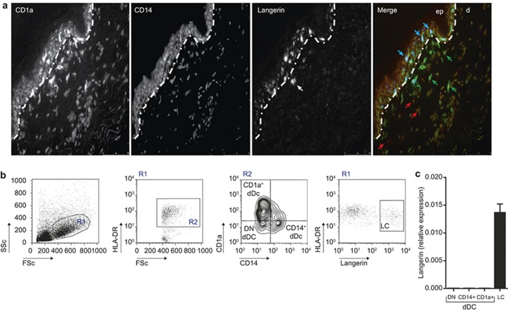
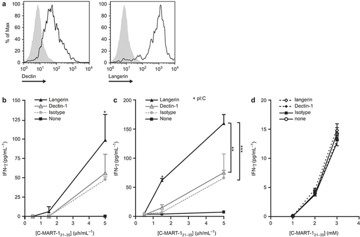
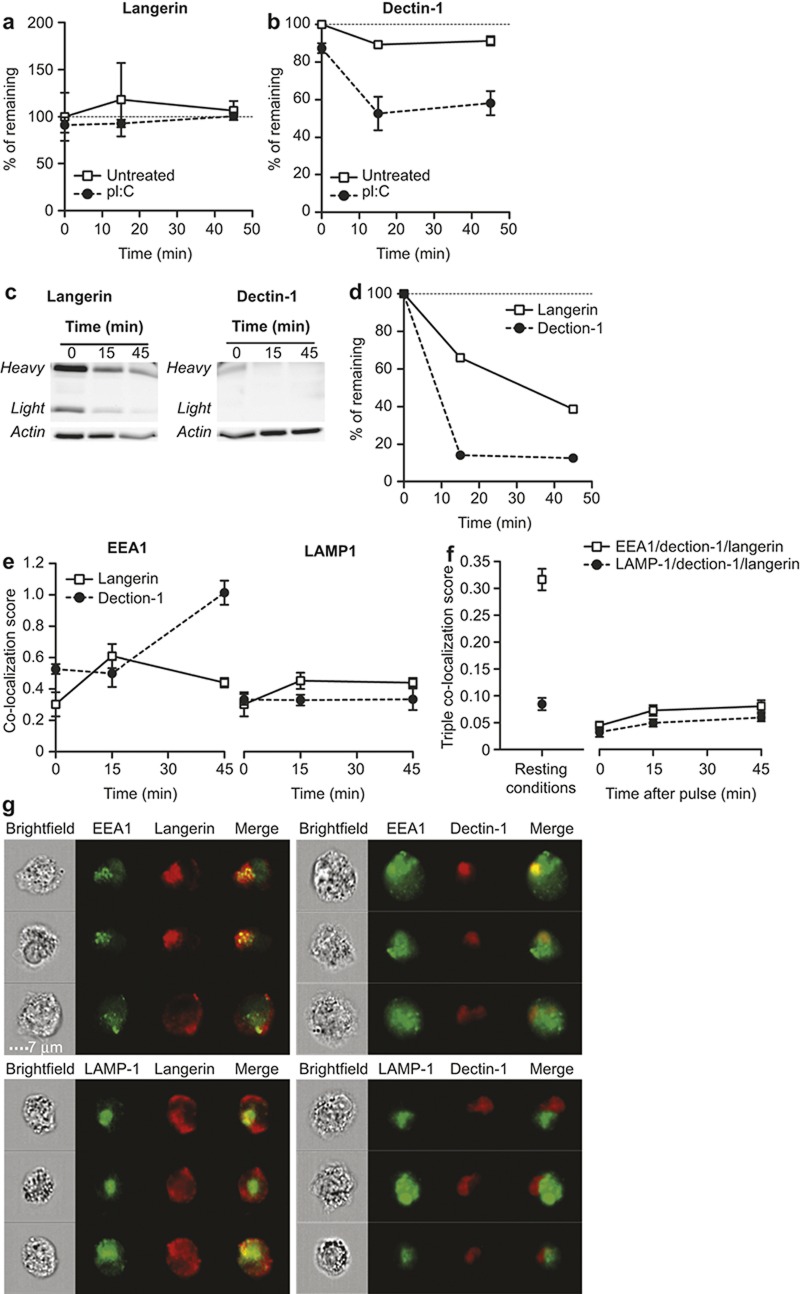
The potential of the skin immune system to generate immune responses is well established, and the skin is actively exploited as a vaccination site. Human skin contains several antigen-presenting cell subsets with specialized functions. In particular, the capacity to cross-present exogenous antigens to CD8+ T cells is of interest for the design of effective immunotherapies against viruses or cancer. Here, we show that primary human Langerhans cells (LCs) were able to cross-present a synthetic long peptide (SLP) to CD8+ T cells. In addition, modification of this SLP using antibodies against the receptor langerin, but not dectin-1, further enhanced the cross-presenting capacity of LCs through routing of internalized antigens to less proteolytic early endosome antigen 1+ early endosomes. The potency of LCs to enhance CD8+ T-cell responses could be further increased through activation of LCs with the toll-like receptor 3 ligand polyinosinic:polycytidylic acid (pI:C). Altogether, the data provide evidence that human LCs are able to cross-present antigens after langerin-mediated internalization. Furthermore, the potential for antigen modification to target LCs specifically provides a rationale for generating effective anti-tumor or anti-viral cytotoxic T lymphocyte responses.


Figures

Figure 1
Figure 1
Langerin is exclusively expressed by human LCs. (a) Staining of a section of steady-state human skin for langerin (blue), CD1a (green), CD14 (red), and Hoechst (yellow), and analysis by fluorescence microscopy. (b) Gating strategy for FACS-sorted LCs, CD14+, CD1a+, and double-negative dermal DCs. (c) Langerin mRNA is exclusively expressed in primary, FACS-sorted LCs and not by the other skin DC subsets. N = 3; each experiment contained sorted cells from at least five skin donors. mRNA values are normalized to GAPDH levels.
Figure 2
Figure 2
Maturation of LCs in vitro upon stimulation with the TLR3 ligand pI:C. (a) Phenotypic characterization of human LCs after 16 h of culture in the presence of the indicated TLR ligands. The mean fluorescence intensity is depicted for MHC class I, CD86, and CD70. The data represent the average ± SEM for three independent skin donors. (b) Cytokines produced by human LCs cultured for 16 h in the presence or absence of the indicated TLR ligands. The data represent the average ± SEM for three independent skin donors. *p < 0.05; **p < 0.01; ***p < 0.001.
Figure 3
Figure 3
pI:C stimulation of LCs results in superior antigen cross-presentation. (a) 1 × 10⁵ T2 cells were incubated with a 16 aa MART-1 peptide, the minimal CD8 epitope or no peptide, after which cells were analyzed for surface HLA-A2 expression by flow cytometry. The data represent the average of three experiments ± SD. **p < 0.01. (b) In total, 2 × 10⁴ HLA-A2+ LCs were incubated with a 16 aa MART-1 peptide, the minimal CD8 epitope or no peptide for 30 min, washed and co-cultured with 1 × 10⁵ MART-1-specific CD8+ T cells. After 24 h of co-culture, T-cell activation was measured by IFN-γ ELISA analysis of the supernatants. The data represent the average of two experiments ± SD. **p < 0.01. (c). Human LCs were incubated with the synthetic long MART-1 peptide and indicated TLR ligands for 3 h, washed and co-cultured with a MART-1-specific CD8+ T-cell clone. After 24 h of co-culture, T-cell activation was measured by IFN-γ ELISA analysis of the supernatants. The data from one representative experiment measured in triplicate is shown, n = 3. *p < 0.05.
Figure 4
Figure 4
SLP conjugated to langerin results in enhanced cross-presentation to antigen-specific CD8+ T cells. (a) Dectin-1 and langerin are highly expressed by human LCs as measured by flow cytometry. Open histograms: specific antibody; filled histograms: isotype control. The data are representative of three donors. (b and c) MART-1 (C-YTTAEELAGIGILTV) peptide was conjugated to anti-langerin and anti-dectin-1 or mIgG1 isotype control mAbs, incubated with human LCs for 3 h in the presence (c), or absence (b) of 20 μg mL–1 pI:C and co-cultured with an MART-1-specific CD8+ T-cell clone. The activation of the T cells was measured by IFN-γ ELISA analysis of the supernatants taken after 24 h of co-culture. The data are representative of two independent experiments and depict the average ± SEM of triplicates. (d) Human LCs were incubated with MART-1 (C-YTTAEELAGIGILTV) peptide in the presence or absence of anti-langerin, anti-dectin-1, or 20 μg mL–1 pI:C for 3 h, and then washed and co-cultured with an MART-1-specific CD8+ T-cell clone. The activation of the T cells was measured by IFN-γ ELISA analysis of the supernatants taken after 24 h of co-culture.
Figure 5
Figure 5
Langerin routes to less degradative endosomal compartments in LCs after antigenic pulse compared to dectin-1 A+B. The remaining fluorescence of anti-langerin (a) and anti-dectin-1 antibodies (b) in a pulse–chase in the presence or absence of pI:C. C+D. Pulse–chase of anti-langerin and anti-dectin-1 antibody degradation in LCs by western blotting using an anti-mouse Fc peroxidase-labeled antibody. Staining is shown in (c), and band intensity quantification in (d). (e) Co-localization of langerin and dectin-1 with the intracellular compartments EEA-1 and LAMP-1 after receptor binding and internalization at the cell surface. (f) Three-color co-localization score of langerin, dectin-1 and EEA-1, or LAMP-1 in resting conditions (left panel) or after receptor triggering at the cell surface (right panel) measured by imaging flow cytometry (representative images are shown in Supplementary Figure S4). (g) Randomly selected LCs representing the data depicted in e (15-min time point).

References

    1. Heath WR, Belz GT, Behrens GM, Smith CM, Forehan SP, Parish IA et al. Cross-presentation, dendritic cell subsets, and the generation of immunity to cellular antigens. Immunol Rev 2004; 199: 9–26. - PubMed
    1. Joffre OP, Segura E, Savina A, Amigorena S. Cross-presentation by dendritic cells. Nat Rev Immunol 2012; 12: 557–569. - PubMed
    1. Valladeau J, Dezutter-Dambuyant C, Saeland S. Langerin/CD207 sheds light on formation of birbeck granules and their possible function in Langerhans cells. Immunol Res 2003; 28: 93–107. - PubMed
    1. Feinberg H, Taylor ME, Razi N, McBride R, Knirel YA, Graham SA et al. Structural basis for langerin recognition of diverse pathogen and mammalian glycans through a single binding site. J Mol Biol 2011; 405: 1027–1039. - PMC - PubMed
    1. Furio L, Briotet I, Journeaux A, Billard H, Peguet-Navarro J. Human Langerhans cells are more efficient than CD14(-)CD1c(+) dermal dendritic cells at priming naive CD4(+) T cells. J Invest Dermatol 2010; 130: 1345–1354. - PubMed

MeSH terms