. 2015 Dec 1;87(23):11660-5.
doi: 10.1021/acs.analchem.5b00702. Epub 2015 Nov 11.

Aptamer-Phage Reporters for Ultrasensitive Lateral Flow Assays

Affiliations

Aptamer-Phage Reporters for Ultrasensitive Lateral Flow Assays

Meena Adhikari et al. Anal Chem. .

Abstract

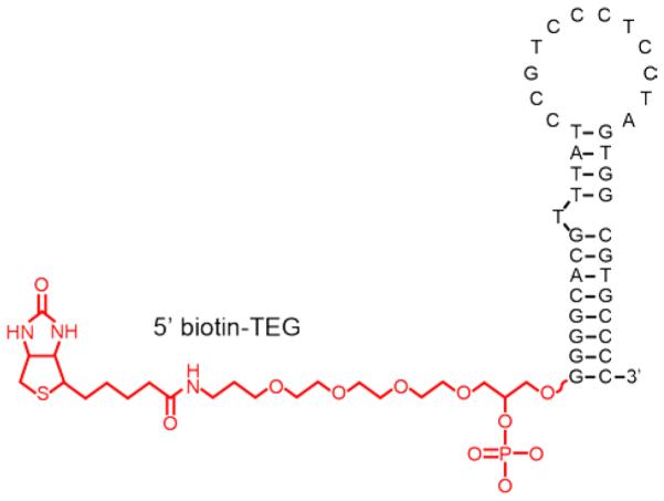
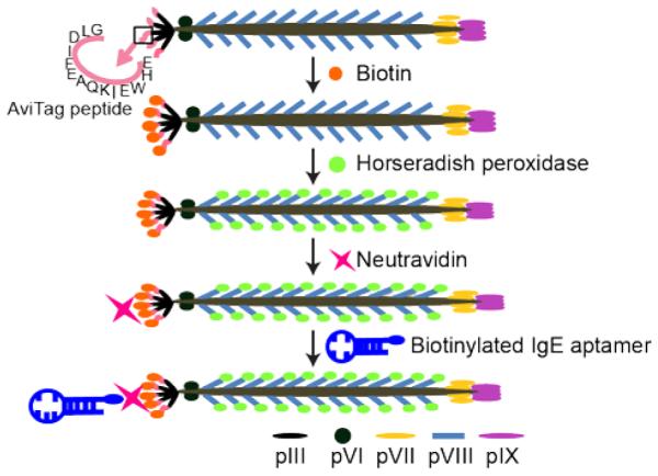
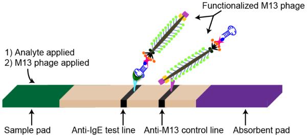
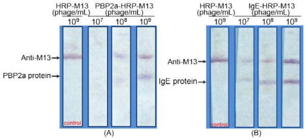
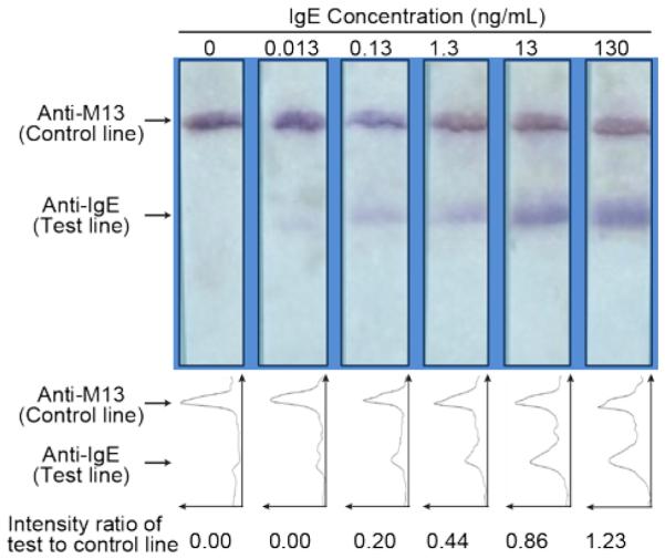
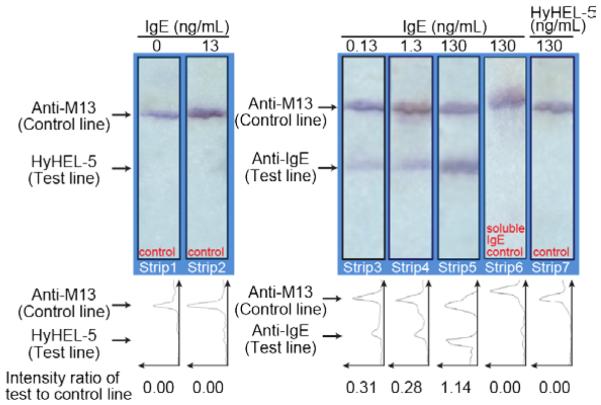
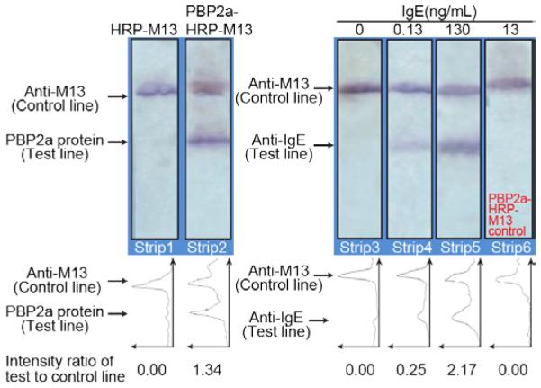
We introduce the modification of bacteriophage particles with aptamers for use as bioanalytical reporters, and demonstrate the use of these particles in ultrasensitive lateral flow assays. M13 phage displaying an in vivo biotinylatable peptide (AviTag) genetically fused to the phage tail protein pIII were used as reporter particle scaffolds, with biotinylated aptamers attached via avidin-biotin linkages, and horseradish peroxidase (HRP) reporter enzymes covalently attached to the pVIII coat protein. These modified viral nanoparticles were used in immunochromatographic sandwich assays for the direct detection of IgE and of the penicillin-binding protein from Staphylococcus aureus (PBP2a). We also developed an additional lateral flow assay for IgE, in which the analyte is sandwiched between immobilized anti-IgE antibodies and aptamer-bearing reporter phage modified with HRP. The limit of detection of this LFA was 0.13 ng/mL IgE, ∼100 times lower than those of previously reported IgE assays.


Figures

Figure 1
Figure 1
Biotin-TEG IgE aptamer (secondary structure adapted from Reference 29).
Figure 2
Figure 2
Functionalization of M13 phage with IgE aptamers and Horseradish peroxidase (HRP). Phage displaying AviTag peptides are biotinylated using Biotin ligase. Biotinylated phage are covalently modified with HRP on the major coat protein pVIII before neutravidin is bound to biotinylated AviTag on HRP-labeled phage. Neutravidin-functionalized phage are then conjugated with the biotinylated IgE aptamer.
Figure 3
Figure 3
Aptamer-phage lateral flow assay. The M13 phage SAM-AviTag protein, pIII, was specifically biotinylated and functionalized with analyte-specific biotinylated aptamers, and the M13 coat proteins were functionalized with Horseradish peroxidase. The detection line contains IgE-specific antibodies, and the control line has anti-M13 antibodies. Captured HRP-labeled phage were detected on the test and control lines using the chromogenic substrate, 3’,3,5,5’-tetramethylbenzidine (TMB).
Figure 4
Figure 4
Direct detection of spotted target analytes (PBP2a (A) or IgE (B)) with aptamer-phage. (A) PBP2a protein and anti-M13 antibodies were spotted on the test and control lines, respectively. (B) IgE protein and anti-M13 antibodies were spotted on the test and control lines, respectively. Different concentrations of aptamer-phage constructs were then passed through the membranes. Phage bearing HRP reporters but no aptamers (HRP-M13) served as a specificity control.
Figure 5
Figure 5
Lateral flow assay with aptamer-phage for IgE detection. Various concentrations of IgE (volume: 10 μL) were detected using anti-IgE antibodies at the test line and IgE aptamer-HRP-phage reporters. Control line consisted of anti-M13 antibodies. Line intensity profiles as evaluated by ImageJ density analysis of the LFA strips are shown below each strip. The area under each peak was numerically integrated using the ImageJ Gel Analysis Toolbox to give the intensity for that line. The intensity of the test line divided by the intensity of the control line for each strip is shown below each line intensity profile.
Figure 6
Figure 6
Control for non-specific binding of IgE and aptamer-phage to an unrelated protein (the murine anti-lysozyme IgG antibody HyHEL-5). Strips 1 & 2; HyHEL-5 was spotted on the test line and anti-M13 antibodies on the control line. IgE protein (100 μL in LFA buffer) was passed through the membrane and aptamer-phage were added as reporters. No signal was observed on the HyHEL-5 test line. Strips 3, 4 & 5; To confirm specificity and sensitivity of the assay, varied concentrations of IgE were passed through the membrane. Strip 6; Competition assay where free IgE aptamer was passed on the strip prior to offering the aptamer-phage construct; 130 ng/mL IgE in 100 μL of LFA buffer were passed, followed by soluble IgE aptamer (10 μM aptamer in 100 μL of LFA buffer) and then the aptamer-phage construct was offered. No signal was observed on the test line confirming that soluble anti-IgE aptamer competes with aptamer-phage for the available binding sites on the IgE protein. Strip 7; 100 μL of HyHEL-5 in LFA buffer was passed through the membrane with anti-IgE and anti-M13 lines and detected using the IgE aptamer-phage reporter. No signal was observed on the HyHEL-5 test line, confirming specificity of the assay. Line intensity profiles as evaluated by ImageJ density analysis of the LFA strips. The area under each peak was numerically integrated using the ImageJ Gel Analysis Toolbox to give the intensity for that line. The intensity of the test line divided by the intensity of the control line for each strip is shown below each line intensity profile.
Figure 7
Figure 7
PBP2a aptamer-phage as a specificity control for IgE-aptamer-phage reporters. Strips 1 & 2; Strips with PBP2a protein and anti-M13 antibodies on the test and control lines, respectively, were made to evaluate the binding of PBP2a-M13 to PBP2a protein. A clear signal on the test line of strip 2 indicates binding of the PBP2a-phage to the PBP2a protein. Strip 1 with HRP-phage devoid of any signal, confirms that phage do not bind non-specifically to the PBP2a protein. Strips 3, 4 & 5 show the various concentrations of IgE protein (100 μL in LFA buffer) that were used. IgE binds on the anti-IgE test line. Signals on the control lines indicate the proper functioning of the assay. Strip 6; A PBP2a-M13 phage construct was passed through a strip with anti-IgE and anti-M13 lines. The anti-PBP2a aptamer on the phage did not bind to the IgE protein on the test line as indicated by the absence of any signal on the test line. Line intensity profiles as plotted by ImageJ density analyses of the LFA strips are shown below each figure. The area under each peak was numerically integrated using the ImageJ Gel Analysis Toolbox, and the average intensity of the test line divided by the intensity of the control line for each strip is also shown below each line intensity profile.

References

    1. Gubala V, Harris LF, Ricco AJ, Tan MX, Williams DE. Anal Chem. 2012;84:487–515. - PubMed
    1. Ngom B, Guo Y, Wang X, Bi D. Anal Bioanal Chem. 2010;397:1113–1135. - PubMed
    1. Anfossi L, Baggiani C, Giovannoli C, D'Arco G, Giraudi G. Anal Bioanal Chem. 2013;405:467–480. - PubMed
    1. Carter DJ, Cary RB. Nucleic Acids Res. 2007;35:e74. - PMC - PubMed
    1. Liu G, Mao X, Phillips JA, Xu H, Tan W, Zeng L. Anal Chem. 2009;81:10013–10018. - PMC - PubMed

Publication types

MeSH terms

LinkOut - more resources