Review
. 2015 Nov;45(6):1183-212, vi.
doi: 10.1016/j.cvsm.2015.06.006.

Clinical Signs and Diagnosis of the Canine Primary Glaucomas

Affiliations
Review

Clinical Signs and Diagnosis of the Canine Primary Glaucomas

Paul E Miller et al. Vet Clin North Am Small Anim Pract. 2015 Nov.

Abstract

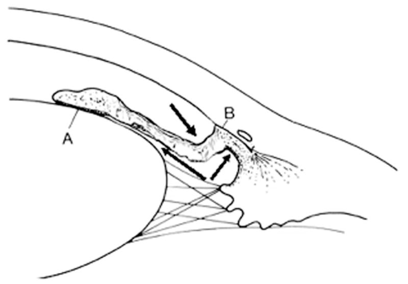
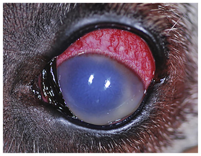
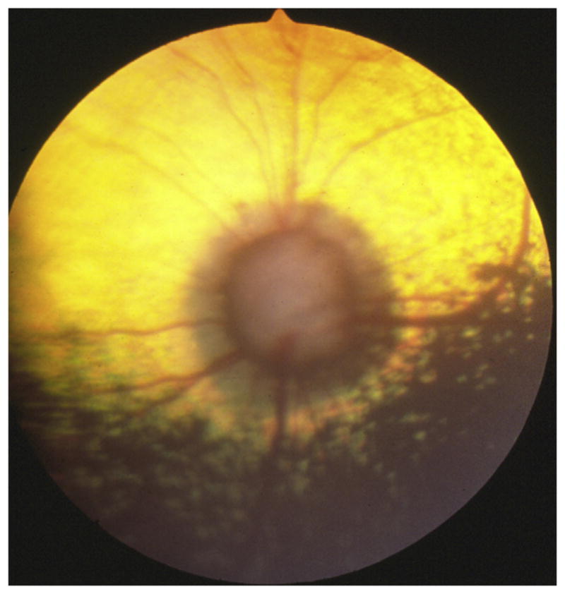
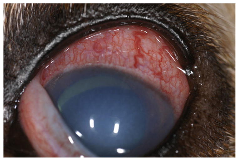
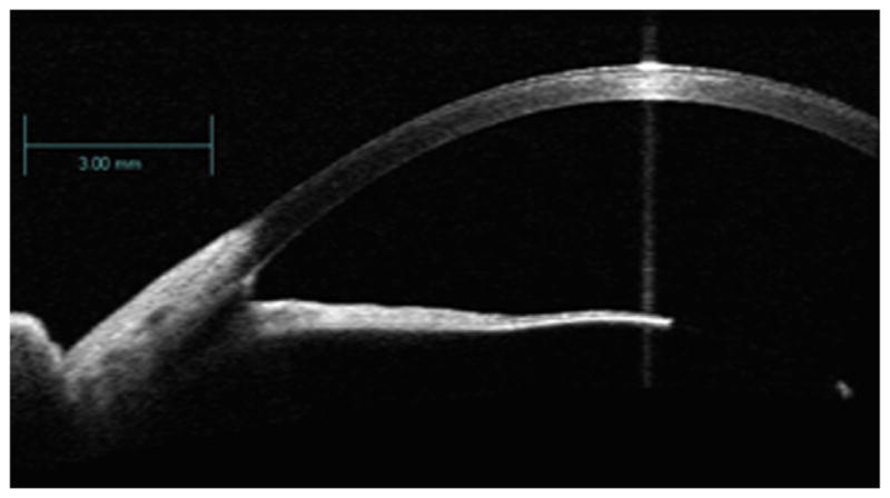
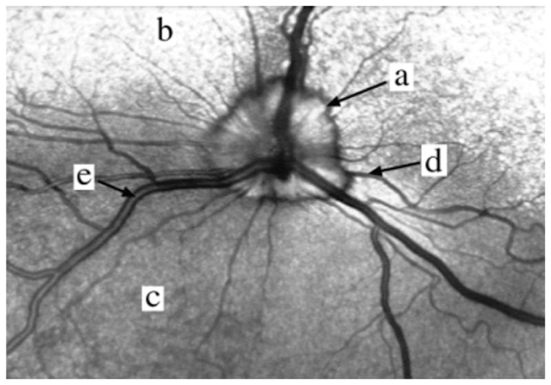
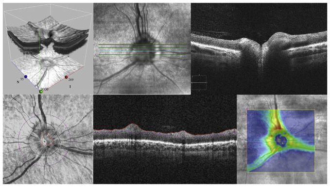
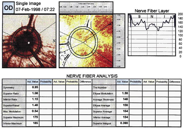
The diagnosis of glaucoma is highly dependent on a working understanding of the clinical signs and available diagnostic procedures. Clinical signs may be attributable to increased intraocular pressure and/or complex alterations in the physiology or molecular biology of the anterior segment, retinal ganglion cells, and optic nerve. Many diagnostic procedures seek to more fully characterize these alterations and to identify which clinical features increase the risk of overt primary angle closure glaucoma (PACG) occurring. Considerable progress has been made in identifying the anatomic features that predispose an eye to PACG, and in elucidating the role of reverse pupillary block.

Keywords: Canine; Glaucoma; Intraocular pressure; Ocular imaging; Optic neuropathy; Tonometry.


Figures

Fig. 1
Fig. 1
Chronic POAG in a beagle. (A) Both pupils are dilated and IOP is increased in both eyes to approximately 50 mm Hg. (B) Fundus photograph of the same dog. The optic disc is depressed from the surface of the fundus (cupped), has little myelin and is darker than normal. The area surrounding the disc has altered reflectivity. (From Miller PE. The glaucomas. In: Maggs DJ, Miller PE, Ofri R, editors. Slatter’s fundamentals of veterinary ophthalmology. 5th edition. St Louis (MO): Elsevier; 2013. p. 259; with permission.)
Fig. 2
Fig. 2
Primary open angle glaucoma in the beagle (same dog as in Fig. 1). (A) The iridocorneal angle is gonioscopically relatively open. (B) High-resolution ultrasound image of the anterior segment. Note that the iris does not have the same configuration as in PACG (see Fig. 3) and that the ciliary cleft is still open (arrow). (From Miller PE. The glaucomas. In: Maggs DJ, Miller PE, Ofri R, editors. Slatter’s fundamentals of veterinary ophthalmology. 5th edition. St Louis (MO): Elsevier; 2013. p. 259; with permission.)
Fig. 3
Fig. 3
Acute PACG in an American cocker spaniel. (A) This disorder first manifests as a unilateral disease, but both eyes are typically ultimately affected. (B) A closed iridocorneal angle on gonioscopy. (C) In the acute stages, the optic disc is pale (poorly perfused) and there is subtle peripapillary edema (arrows). In the latter stages, it looks similar to that seen in Fig. 1. (From Miller PE. The glaucomas. In: Maggs DJ, Miller PE, Ofri R, editors. Slatter’s fundamentals of veterinary ophthalmology. 5th edition. St Louis (MO): Elsevier; 2013. p. 258; with permission.)
Fig. 4
Fig. 4
(A) High-resolution ultrasound image of a normal eye. AC, anterior chamber; C, cornea; CB, ciliary body; I, iris; L, lens. White arrows outline the ciliary cleft. (B) An eye with acute PACG. Note the sigmoidal shape of the iris, increased contact of the peripheral iris with the cornea (black arrow) and collapse of the ciliary cleft (white arrow). (From Miller PE. The glaucomas. In: Maggs DJ, Miller PE, Ofri R, editors. Slatter’s fundamentals of veterinary ophthalmology. 5th edition. St Louis (MO): Elsevier; 2013. p. 258; with permission.)
Fig. 5
Fig. 5
Goniophotograph of a normal dog. A, pupil; B, iris; C, pectinate ligament strands (thin brown lines); D, bluish-white zone of the trabecular meshwork; E, deep pigmented zone; F, superficial pigmented zone; G, cornea. (From Miller PE. The glaucomas. In: Maggs DJ, Miller PE, Ofri R, editors. Slatter’s fundamentals of veterinary ophthalmology. 5th edition. St Louis (MO): Elsevier; 2013. p. 250; with permission.)
Fig. 6
Fig. 6
Marked PLD characterized by broad sheets of tissue. Although IOP was within normal limits, aqueous humor can exit the eye only via a few small “flow holes” (arrows) in the sheets. (From Miller PE. The glaucomas. In: Maggs DJ, Miller PE, Ofri R, editors. Slatter’s fundamentals of veterinary ophthalmology. 5th edition. St Louis (MO): Elsevier; 2013. p. 251; with permission.)
Fig. 7
Fig. 7
Proposed reverse pupillary block theory of the mechanism of PACG in dogs. See text for complete description. PLD holds the peripheral iris in close contact with the peripheral cornea and prevents the angle from opening farther when anterior chamber pressures increase. Stress or excitement puts the pupil into the midrange to dilated position and also increases the choroidal pulse pressure. The increase in choroidal pulse pressure forces small aliquots of aqueous humor into the anterior chamber, which results in slightly higher pressure in the anterior chamber than in the posterior chamber (arrows). This additional fluid cannot escape via the drainage angle and the pressure differential forces the iris into tighter contact with the anterior lens capsule creating pupil block (A), which prevents aqueous from returning to the posterior chamber. Prolonged increases in IOP lead to peripheral anterior synechia (B) and a further impediment to aqueous outflow. (From Miller PE. The glaucomas. In: Maggs DJ, Miller PE, Ofri R, editors. Slatter’s fundamentals of veterinary ophthalmology. 5th edition. St Louis (MO): Elsevier; 2013. p. 260; with permission.)
Fig. 8
Fig. 8
Acute PACG in a great Dane. Notice the marked episcleral vascular congestion, diffuse corneal edema, and midrange to dilated pupil.
Fig. 9
Fig. 9
Optic disc cupping in PACG. Most retinal vessels disappear at the disc edge. The center of the disc is in focus below the level of the retinal surface and is grayish in color. There is also a peripapillary ring of altered retinal reflectivity. (Courtesy of Christopher J. Murphy, DVM, PhD, University of California, Davis, Davis, CA.)
Fig. 10
Fig. 10
Chronic PACG in a dog. The eye is enlarged (buphthalmic), irreversibly blind, and has a subluxated lens.
Fig. 11
Fig. 11
Tonometers commonly used in the diagnosis of PACG. Far left, Schiotz indentation tonometer; middle, Tono-Pen Vet applanation tonometer; far right, TonoVet rebound tonometer.
Fig. 12
Fig. 12
Incorrect technique for opening the eyelids for tonometry. The lids are being held open with pressure on the globe, thereby artificially increasing IOP.
Fig. 13
Fig. 13
Proper technique for holding the eyelids open. The lids are opened from over the boney orbital rim thereby avoiding pressure on the globe.
Fig. 14
Fig. 14
View through a Goldmann 3-mirror gonioprism. The central lens allows for viewing of the fundus with a slit-lamp biomicroscope. The 3 surrounding mirrors have different degrees of angulation so as to facilitate examination of different structures within the irido-corneal angle.
Fig. 15
Fig. 15
Schematic drawing of a grading scheme for the width of the iridocorneal angle. The ratio of the width of the anterior opening of the ciliary cleft (A) and the distance from the origin of the pectinate ligaments to the anterior surface of the cornea (B) is estimated. Also shown are the pupil (C), iris (D), pectinate ligament (E), deep pigmented zone (F), superficial pigmented zone (G), and cornea (H). (From Ekesten B, Narfström K. Correlation of morphologic features of the iridocorneal angle to intraocular pressure in Samoyeds. Am J Vet Res 1991;52:1875; with permission.)
Fig. 16
Fig. 16
AS-OCT image of a dog. This technique uses near-infrared light instead of sound to image the iridocorneal angle. As such, visualization of the ciliary body and lens are not as apparent as that observed on high-resolution ultrasound (see Figs. 2 and 4 for comparison). (Courtesy of Carol Rasmussen, MS, University of Wisconsin-Madison, Madison, WI.)
Fig. 17
Fig. 17
Confocal Scanning Laser Ophthalmoscopy of a dog. A laser is used to image the optic disc (a), tapetum lucidum (b), nontapetal zone (c), retinal arterioles (d), and retina venules (e). (From Rosolen SG, Saint-Macary G, Gautier V, et al. Ocular fundus images with confocal scanning laser ophthalmoscopy in the dog, monkey and minipig. Vet Ophthalmol 2001;4(1):42; with permission.)
Fig. 18
Fig. 18
Spectral-domain OCT image of a normal dog. This instrument provides high-resolution images of the posterior segment and includes an adjustable 3-dimensional view of the optic disc and surrounding retina (upper left), an ophthalmoscopic view of the optic disc denoting where line scans were obtained (upper middle), a cross-sectional view through the optic disc at the selected line scan (upper right), circular scans around the optic disc (lower right), cross-sectional views of the retina (lower middle), and color-coded thickness mapping of the optic disc or retina (lower right). (Courtesy of Carol Rasmussen, MS, University of Wisconsin-Madison, Madison, WI.)
Fig. 19
Fig. 19
Image from a normal eye with a scanning laser polarimeter (Gdx). Images include a fundus image from the scanned area and a color-coded thickness retardation map of the RNFL that shows thinner areas in blue and black and thicker areas in red, orange, and yellow. A variety of printouts are also presented. (From Garcia-Sánchez GA, Gil-Carrasco F, Román JJ, et al. Measurement of the retinal nerve fiber layer thickness in normal and glaucomatous Cocker Spaniels by scanning laser polarimetry. Vet Ophthalmol 2007;10 Suppl 1:81; with permission.)

References

    1. Spaeth GL, Waisbourd M. Definitions: what is glaucoma worldwide? In: Shaarawy TM, Sherwood MB, Hitchings RA, et al., editors. Glaucoma: medical diagnosis & therapy. St Louis (MO): Elsevier Ltd; 2015. pp. 311–24.
    1. Foster PJ, Buhrmann R, Quigley HA, et al. The definition and classification of glaucoma in prevalence surveys. Br J Ophthalmol. 2002;86:238–42. - PMC - PubMed
    1. Ahonen SJ, Pietilä E, Mellersh CS, et al. Genome-wide association study identifies a novel canine glaucoma locus. PLoS One. 2013;8:e70903. - PMC - PubMed
    1. Kuchtey J, Kunkel J, Esson D, et al. Screening ADAMTS10 in dog populations supports Gly661Arg as the glaucoma-causing variant in beagles. Invest Ophthalmol Vis Sci. 2013;54:1881–6. - PMC - PubMed
    1. Kuchtey J, Olson LM, Rinkoski T, et al. Mapping of the disease locus and identification of ADAMTS10 as a candidate gene in a canine model of primary open angle glaucoma. PLoS Genet. 2011;7:e1001306. - PMC - PubMed

Publication types