. 2015 Oct 20;13(3):524-532.
doi: 10.1016/j.celrep.2015.09.014. Epub 2015 Oct 8.

Tankyrase Inhibitors Target YAP by Stabilizing Angiomotin Family Proteins

Affiliations

Tankyrase Inhibitors Target YAP by Stabilizing Angiomotin Family Proteins

Wenqi Wang et al. Cell Rep. .

Abstract

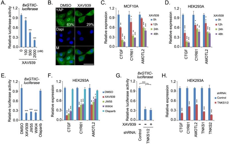
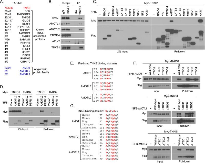
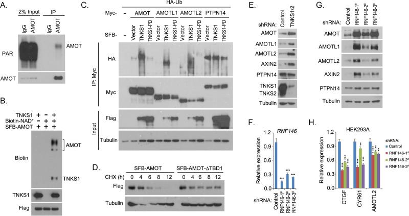
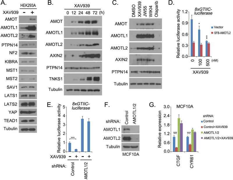
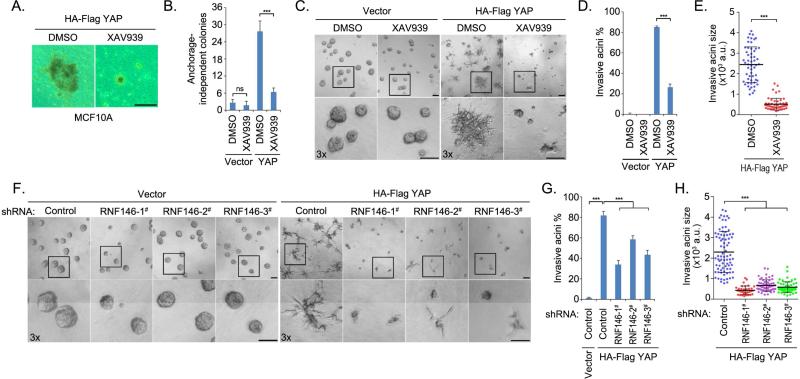
As the key effector in the Hippo pathway, YAP was identified as an oncoprotein whose expression is elevated in various human cancers. However, the development of potentially therapeutic compounds targeting YAP has been slow and limited. Here, we find that tankyrase inhibitors suppress YAP activity. This effect is mediated by anigomotin (AMOT) family proteins. Tankyrases associate with AMOT family proteins and promote their degradation through E3 ligase RNF146. By antagonizing tankyrase activity, tankyrase inhibitors stabilize AMOT family proteins, thereby suppressing YAP oncogenic functions. Together, our studies not only demonstrate the tankyrase-RNF146-AMOT axis as an upstream pathway regulating YAP but also reveal a therapeutic opportunity in targeting YAP for cancer treatment.


Figures

Figure 1
Figure 1. Tankyrase inhibitors suppressed YAP activity
(A) XAV939 suppressed YAP/TEAD luciferase reporter activity. Luciferase reporter assay was performed in HeLa cells treated with XAV939 at indicated concentrations for 24 h. pSV40-Renilla was used as internal control. (B) XAV939 treatment partially translocated YAP from nucleus into cytoplasm. Low-density MCF10A cells were treated with dimethyl solfoxide (DMSO) or XAV939 (10 μM) for 48 h, and the percentages of nuclear YAP are shown. Nucleus is indicated by DAPI staining. M, merged. Scale bar, 20μm. (C-D) XAV939 treatment suppressed YAP transcriptional activity. The transcripts of YAP target genes were detected by quantitative PCR in MCF10A (C) and HEK293A (D) cells. Cells were treated with XAV939 (10 μM) for the indicated time periods. (E-F) Tankyrase inhibitors suppressed YAP activities. YAP/TEAD-luciferase reporter activity (E) and the transcripts of YAP target genes (F) were detected in cells treated with indicated inhibitors (10 μM) for 24h. (G-H) Double knockdown of tankyrase 1 and 2 suppressed YAP activities. YAP/TEAD-luciferase reporter activity (G) and the transcripts of YAP target genes (H) were detected in cells transduced with indicated shRNAs. For the luciferase reporter assay, HeLa cells were treated with XAV939 (10 μM) for 24 h. For all panels, * p<0.05, ** p<0.01 and *** p<0.001.
Figure 2
Figure 2. Tankyrases associated with angiomotin family proteins
(A) Association of angiomotin family proteins with tankyrase 1 was identified by tandem affinity purification–mass spectrometry (TAP-MS) in HEK293T cells. Bait protein is marked in red. Identified angiomotin family proteins AMOT, AMOTL1, and AMOTL2 are marked in blue. The left row of numbers represents unique peptide number/total peptide number. The known tankyrase 1 (TNKS1)–associated proteins are indicated. (B) TNKS1 interacted with AMOT proteins. Endogenous immunoprecipitation (IP) was performed using extract prepared from HEK293T cells; normal rabbit IgG was taken as control. (C) TNKS1 specifically associated with AMOT in the Hippo pathway. Myc-tagged TNKS1 was co-expressed with indicated SFB-tagged proteins in HEK293T cells and cell lysates were subjected to pulldown assays. (D) Both TNKS1 and TNKS2 associated with AMOTL2. Myc-tagged TNKS1 or TNKS2 was co-expressed with indicated SFB-tagged proteins in HEK293T cells, and cell lysates were subjected to pulldown assays. (E) The predicted tankyrase-binding domains (TBD) for AMOT family proteins are listed. The key amino acids for each TBD are indicated in red. (F) The first predicted TBD was identified as the bona-fide TBD for each AMOT proteins. Myc-tagged TNKS1 was co-expressed with indicated SFB-tagged AMOT proteins or their TBD-deleted mutants in HEK293T cells, and cell lysates were subjected to pulldown assays. (G) The sequence alignments for the TBD in AMOT family proteins from different species are shown. The key amino acids for each TBD are indicated in red.
Figure 3
Figure 3. Tankyrase-RNF146 axis promoted the degradation of AMOT proteins
(A) AMOT protein was ribosylated in vivo. Endogenous immunoprecipitation (IP) was performed using extract prepared from HEK293T cells containing ADP-HPD (5 μM); normal rabbit IgG was taken as control. PAR, poly-ADP ribosylation polymer antibody. (B) Tankyrase 1 (TNKS1) promoted ribosylation of AMOT protein in vitro. In vitro ribosylation assay was performed and the ribosylated proteins were indicated by the incorporation of biotinylated NAD+ and detected by anti-biotin antibody. (C) Tankyrase 1 (TNKS1) induced the ubiquitination of AMOT proteins. HA-tagged ubiquitin (Ub) was co-expressed with indicated proteins in HEK293T cells for 24 h. Cells were treated with proteasome inhibitor MG132 (10 μM) for 6 h and subjected to immunoprecipitation (IP) assay. TNKS1-PD is the inactive mutant for TNKS1. (D) Tankyrase-binding domain (TBD) deletion mutation stabilized the AMOT protein. AMOT or its TBD-deleted mutant (AMOT-ΔTBD1) was expressed in HEK293T cells for 24 h. Cells were treated with cycloheximide (CHX; 100 μg/mL) and collected at different time points for Western blotting. (E) Loss of tankyrase 1/2 stabilized AMOT proteins. Indicated proteins were detected in control and tankyrase 1/2 knockdown (shRNA-transduced) HEK293A cells by Western blotting. (F-G) Loss of RNF146 stabilized AMOT proteins. RNF146 knockdown efficiency was shown by quantitative PCR (F). Indicated proteins were detected in control and three groups of RNF146 shRNA-transduced HEK293A cells by Western blotting (G). (H) Loss of RNF146 suppressed the transcripts of YAP target genes. The transcripts were detected by quantitative PCR in control and three groups of RNF146 shRNA-transduced HEK293A cells. For panels F and H, ** p<0.01 and *** p<0.001.
Figure 4
Figure 4. Tankyrase inhibitors targeted YAP through AMOT family proteins
(A) XAV939 treatment specifically increased the levels of AMOT family proteins in the Hippo pathway. Western blot was performed in dimethyl sulfoxide (control)– and XAV939– treated HEK293A cells with indicated antibodies. (B) XAV939 treatment increased the levels of AMOT family proteins in a time-dependent manner. HEK293A cells treated with XAV939 (10 μM) were collected at indicated time points for Western blotting with indicated antibodies. (C) AMOT proteins were stabilized by treatment with tankyrase inhibitors. HEK293A cells treated with the indicated inhibitors (10 μM) for 24 h were subjected to Western blotting with indicated antibodies. (D) Overexpression of AMOTL2 attenuated XAV939-induced suppression of YAP. YAP/TEAD luciferase reporter assay was performed in HeLa cells transfected with vector or SFB-AMOTL2 and treated with indicated doses of XAV939 for 24 h. pSV40-Renilla was used as internal control. (E-G) Loss of AMOTL1/2 attenuated XAV939-induced suppression of YAP. Control and AMOTL1/2 shRNA-transduced HeLa and MCF10A cells were subjected to YAP/TEAD luciferase reporter assay (E) and transcription analysis of YAP target genes by quantitative PCR (G), respectively. The downregulation of AMOTL1 and AMOTL2 in MCF10A cells is shown (F). Both HeLa and MCF10A cells were pre-treated with XAV939 (10μM) for 24 h before collection. For panels D, E, and G, * p<0.05 and *** p<0.001.
Figure 5
Figure 5. Tankyrase inhibitors targeted the oncogenic functions of YAP
(A-B) XAV939 treatment suppressed the anchorage-independent growth of YAP-transformed MCF10A cells. Vector- or YAP-overexpressing MCF10A cells were subjected to soft agar assay and treated dimethyl sulfoxide (DMSO) or XAV939 (30 μM) for 4 weeks. Representative colonies are shown (A) and were quantified (B). Data are presented as means ± standard deviation (s.d.) from three independent experiments. (C-E) XAV939 treatment inhibited YAP-induced MCF10A invasive acini formation. Vector- or YAP-overexpressing MCF10A cells were subjected to 3D culture in matrigel and were treated with DMSO or XAV939 (30 μM) for 5 days. Representative acini are shown (C). The percentage (D) and the size (E) of invasive acini are quantified. Data are presented as means ± s.d. from three independent experiments. a.u., arbitrary unit. (F-H) Loss of RNF146 suppressed YAP-induced MCF10A invasive acini formation. Vector- or YAP-overexpressing MCF10A cells transduced with control or one of three RNF146 shRNAs were subjected to 3D culture in matrigel and treated with DMSO or XAV939 (30 μM) for 5 days. Representative acini (F) and quantification of percentage (G) and size (H) of invasive acini are shown. Data are presented as means ± s.d. from three independent experiments. a.u., arbitrary unit. For all the panels, ns, not significant; *** p<0.001; Scale bar, 100μm.

References

    1. Adler JJ, Johnson DE, Heller BL, Bringman LR, Ranahan WP, Conwell MD, Sun Y, Hudmon A, Wells CD. Serum deprivation inhibits the transcriptional co-activator YAP and cell growth via phosphorylation of the 130-kDa isoform of Angiomotin by the LATS1/2 protein kinases. Proc Natl Acad Sci U S A. 2013;110:17368–17373. - PMC - PubMed
    1. Camargo FD, Gokhale S, Johnnidis JB, Fu D, Bell GW, Jaenisch R, Brummelkamp TR. YAP1 increases organ size and expands undifferentiated progenitor cells. Curr Biol. 2007;17:2054–2060. - PubMed
    1. Chan SW, Lim CJ, Chong YF, Pobbati AV, Huang C, Hong W. Hippo pathway-independent restriction of TAZ and YAP by angiomotin. J Biol Chem. 2011;286:7018–7026. - PMC - PubMed
    1. Chan SW, Lim CJ, Guo F, Tan I, Leung T, Hong W. Actin-binding and cell proliferation activities of angiomotin family members are regulated by Hippo pathway-mediated phosphorylation. J Biol Chem. 2013;288:37296–37307. - PMC - PubMed
    1. Dai X, She P, Chi F, Feng Y, Liu H, Jin D, Zhao Y, Guo X, Jiang D, Guan KL, et al. Phosphorylation of angiomotin by Lats1/2 kinases inhibits F-actin binding, cell migration, and angiogenesis. J Biol Chem. 2013;288:34041–34051. - PMC - PubMed

Publication types

MeSH terms