. 2015 Oct 12:6:8368.
doi: 10.1038/ncomms9368.

Ape parasite origins of human malaria virulence genes

Affiliations

Ape parasite origins of human malaria virulence genes

Daniel B Larremore et al. Nat Commun. .

Abstract

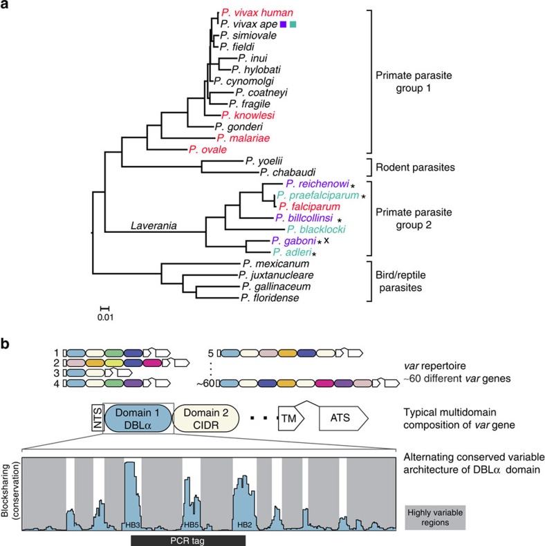
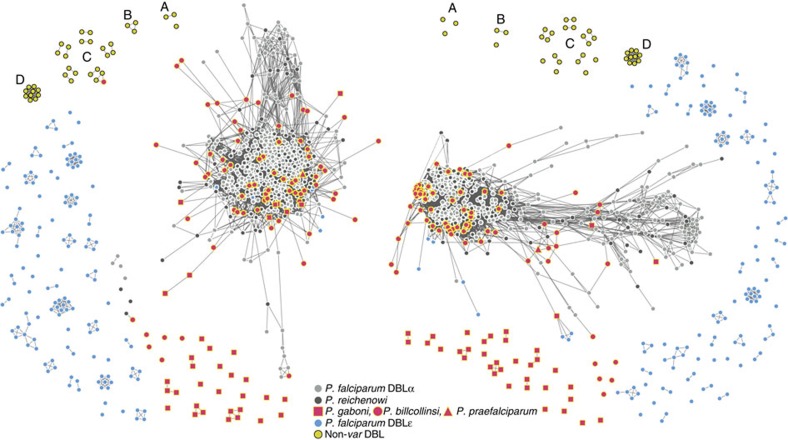
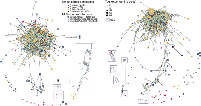
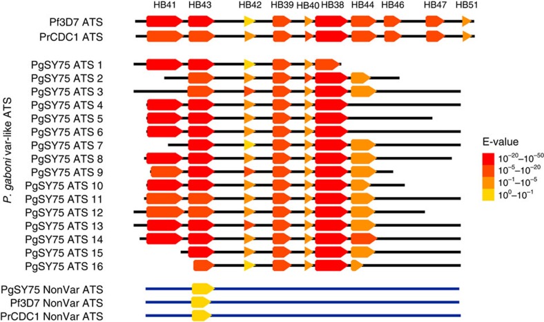
Antigens encoded by the var gene family are major virulence factors of the human malaria parasite Plasmodium falciparum, exhibiting enormous intra- and interstrain diversity. Here we use network analysis to show that var architecture and mosaicism are conserved at multiple levels across the Laverania subgenus, based on var-like sequences from eight single-species and three multi-species Plasmodium infections of wild-living or sanctuary African apes. Using select whole-genome amplification, we also find evidence of multi-domain var structure and synteny in Plasmodium gaboni, one of the ape Laverania species most distantly related to P. falciparum, as well as a new class of Duffy-binding-like domains. These findings indicate that the modular genetic architecture and sequence diversity underlying var-mediated host-parasite interactions evolved before the radiation of the Laverania subgenus, long before the emergence of P. falciparum.


Figures

Figure 1
Figure 1. Characterization of Laverania var gene sequences.
(a) Phylogeny of Plasmodium species. The tree was constructed from mitochondrial sequences (2.4-kb spanning cox1 and cytB). The scale bar indicates 0.01 substitutions per site. Colours indicate species infecting humans (red), chimpanzees (purple) and gorillas (aqua). Asterisks indicate successful PCR amplification of var sequences; a cross indicates identification of var-like genes in near-full-length P. gaboni genomes. (b) Three-level schematic of modular var diversity, structure and architecture. Coloured ovals represent classes of DBL or CIDR domains. White boxes represent the N-terminal segment (NTS), transmembrane (TM) and acidic terminal segment (ATS) domains; a wedge between TM and ATS domains indicates the intron that separates the two var exons. Alternating conserved variable architecture is illustrated using blocksharing (see the Methods section) between one representative DBLα domain (DD2var11) and other DBLα domains published by Rask et al. A black bar indicates the location of the PCR amplified DBLα tag region, which spans three conserved homology blocks (HB3, HB5 and HB2), 72–147 amino acids in length.
Figure 2
Figure 2. Networks of DBLα sequences from P. reichenowi and P. falciparum.
Each node represents a DBLα HVR sequence and each link represents a shared amino-acid substring of significant length. Laverania species and strain origin is indicated by node colour and shape. Left and right networks correspond to left and right HVRs, respectively. P. falciparum and P. reichenowi sequences do not cluster by species or sample in either HVR. Link lengths and node placements are determined by a force-directed layout to better reveal structure, if it exists (see the Methods section). Additional analyses of these networks are shown in Supplementary Fig. 1.
Figure 3
Figure 3. Networks of DBL sequences from Laverania single-species infections in the context of known DBLα and non-DBLα sequences.
Each node represents a DBL HVR sequence from a single-species infection and each link represents a shared amino-acid substring of significant length. Note that for each sample, only unique var DBL haplotypes were included in the network analysis. Nodes with zero links indicate sequences that share no significant amino-acid substrings with other sequences. Networks were built separately for each HVR, where mosaic diversity is highest (see the Methods section). Colours correspond to Laverania species as indicated; annotated yellow nodes correspond to (A) dblsmsp1 and (B) dblmsp2 from Pf3D7, PfIT and PrCDC; (C) both DBL domains from ebl1, eba140, eba165, eba175 and eba181 of Pf3D7 and PfIT; (D) P. vivax Duffy-binding proteins; see Supplementary Table 3 for a comprehensive list of non-DBLα sequences.
Figure 4
Figure 4. Networks of DBL sequences from single- and multi-species Laverania infections.
Each node represents a DBL HVR sequence and each link represents a shared amino-acid substring of significant length. Note that for each sample only unique var DBL haplotypes were included in the network analysis. Nodes with zero links indicate sequences that share no significant amino-acid substrings with other sequences. Networks were built separately for each HVR, where mosaic diversity is highest (see the Methods section). Circular nodes represent chimpanzee parasites and square nodes represent gorilla parasites. Node colour corresponds to species and node size corresponds to tag length as indicated. DBLx sequences are enclosed in boxes. Annotations call attention to (A) P. praefalciparum single-species infection sequence; (B) DBLx sequences from gorilla samples, hypothesized to be P. adleri, that share mosaic elements with DBLx chimpanzee parasites.
Figure 5
Figure 5. Conservation of var ATS domain homology block structure in P. gaboni.
The homology block (HB) structure of var ATS domains identified in 16 contigs of a near complete P. gaboni genome (PgSY75) are shown in relation to representative P. falciparum and P. reichenowi var ATS domains (Pf3D7 and PrCDC1, top) as well as a non-var “ATS-like” domain of the P. falciparum PF3D7_0113800 gene and its P. reichenowi and P. gaboni orthologues (bottom). HBs (arrows) were predicted by VarDom 1.0 and annotated in an alignment of all 20 sequences. Colours correspond to VarDom reported E-values, representing an estimate of the likelihood of observing such a match by random chance. Black lines indicate the relative length of each sequence.
Figure 6
Figure 6. Shared synteny of var-like genes in P. falciparum, P. reichenowi and P. gaboni.
An open-reading frame (ORF) located downstream of a predicted var-like gene in P. gaboni showed 88% sequence identity (dark grey bars) with a single-copy gene present in both P. falciparum 3D7 (PF3D7_0323800) and P. reichenowi CDC1 (PRCDC_0323100). The P. gaboni var-like gene is syntenic with a var exon 2 pseudogene in both P. falciparum and P. reichenowi, suggesting that a var gene was present at this location in the ancestor of all three Laverania species.

References

    1. Liu W. et al. Origin of the human malaria parasite Plasmodium falciparum in gorillas. Nature 467, 420–425 (2010). - PMC - PubMed
    1. Reichenow E. Über das vorkommen der malariaparasiten des menschen bei den afrikanischen menschenaffen. Centralbl. f. Bakt. I. Abt. Orig 85, 207–216 (1920).
    1. Blacklock B. & Adler S. A parasite resembling Plasmodium falciparum in a chimpanzee. Ann. Trop. Med. Parasitol. 16, 99–107 (1922).
    1. Ollomo B. et al. A new malaria agent in African hominids. PLoS Pathog. 5, e1000446 (2009). - PMC - PubMed
    1. Rich S. M. et al. The origin of malignant malaria. Proc. Natl Acad. Sci. USA 106, 14902–14907 (2009). - PMC - PubMed

Publication types

Substances

Associated data