. 2015 Oct 12;10(10):e0137905.
doi: 10.1371/journal.pone.0137905. eCollection 2015.

Impact of MR Acquisition Parameters on DTI Scalar Indexes: A Tractography Based Approach

Affiliations

Impact of MR Acquisition Parameters on DTI Scalar Indexes: A Tractography Based Approach

Gonzalo Barrio-Arranz et al. PLoS One. .

Abstract

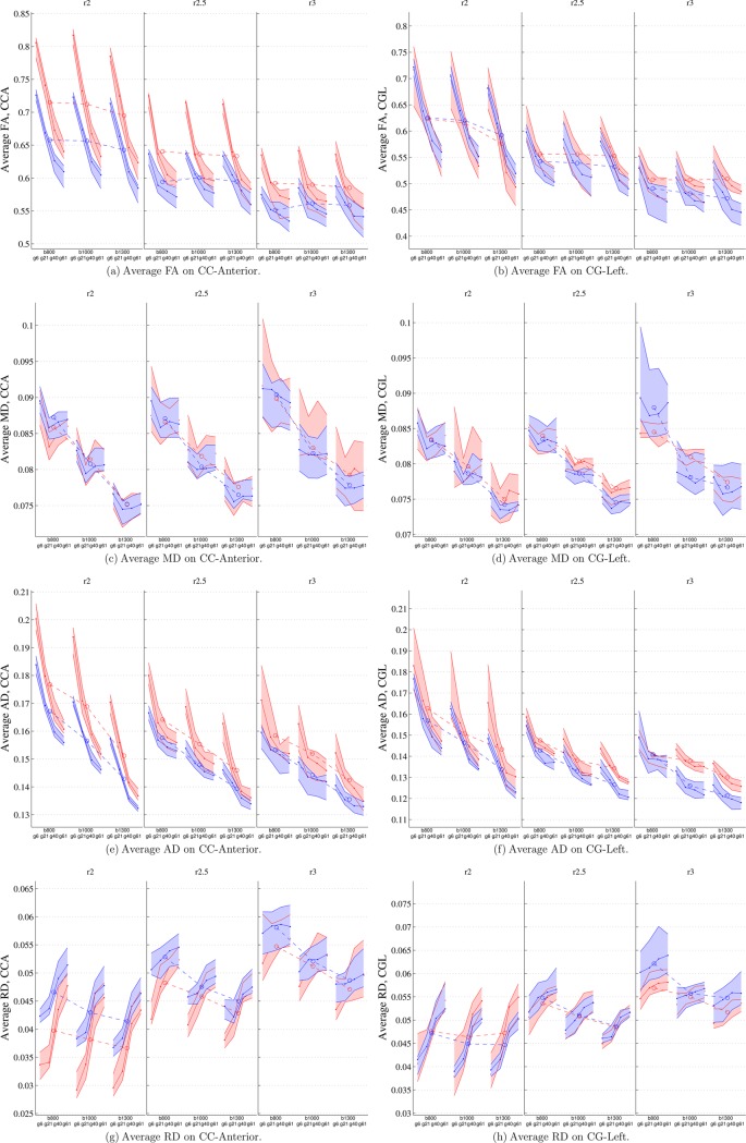
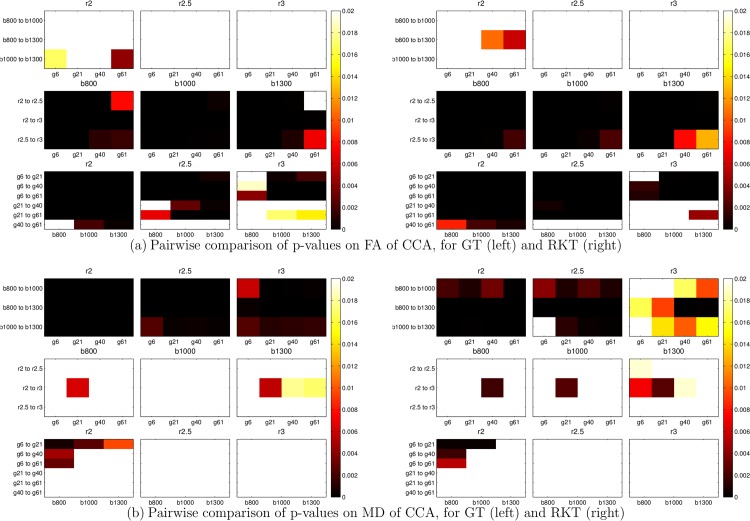
Acquisition parameters play a crucial role in Diffusion Tensor Imaging (DTI), as they have a major impact on the values of scalar measures such as Fractional Anisotropy (FA) or Mean Diffusivity (MD) that are usually the focus of clinical studies based on white matter analysis. This paper presents an analysis on the impact of the variation of several acquisition parameters on these scalar measures with a novel double focus. First, a tractography-based approach is employed, motivated by the significant number of clinical studies that are carried out using this technique. Second, the consequences of simultaneous changes in multiple parameters are analyzed: number of gradient directions, b-value and voxel resolution. Results indicate that the FA is most affected by changes in the number of gradients and voxel resolution, while MD is specially influenced by variations in the b-value. Even if the choice of a tractography algorithm has an effect on the numerical values of the final scalar measures, the evolution of these measures when acquisition parameters are modified is parallel.


Conflict of interest statement

Competing Interests: The authors have declared that no competing interests exist.

Figures

Fig 1
Fig 1. RKT (left) and GT (right) reconstructions of one side of the brain (CC, red and orange; CG, green) for G = 61 gradients, R = 2 × 2 × 2 mm 3, b = 1000 s/mm 2.
Fig 2
Fig 2. Average fiber length L¯ and number of fibers for the RKT (red) and the GT (blue) reconstructions for r = 2mm 3 and b = 1000 s/mm 2.
White boxes stand for G = 6 gradient directions, while solid boxes stand for G = 61. Six different bundles are considered.
Fig 3
Fig 3. 2-D boxplot of Total Volume, VB, (mm 3) versus OV (percentage) for RKT (red) and GT (blue) on CCA and CGL fiber bundles at B = 800 s/mm 2, and different configurations of R and G.
The limits of each rectangle indicate the first and third quartile values for both boxplots. Dotted lines denote median values of each boxplot, while solid and dashed lines gather reconstructions that share the same voxel resolution.
Fig 4
Fig 4. Average values for RKT(red) and GT(blue) on CCA(left column) and CGL (right column) for FA, MD, AD and RD.
Central lines represent median values; upper and lower lines of the shaded areas represent the first and third quartile of the data. Dashed lines between groups represent average values of each group.
Fig 5
Fig 5. Pairwise comparison of p-values.
Black values indicate the existence of a statistical significative difference (p < 0.01) between each pair of acq. parameters.

Similar articles

Cited by

References

    1. Basser PJ, Pierpaoli C. Microstructural and physiological features of tissues elucidated by quantitative-diffusion-tensor MRI. mrm. 1996;111(3):209–219. - PubMed
    1. Horsfield M, Jones D. Applications of DW and DTMRI to white matter diseases—A review. NMR Biomed. 2002;15:570–577. 10.1002/nbm.787 - DOI - PubMed
    1. Salmenpera TM, Simister RJ, Bartlett P, Symms MR, Boulby PA, Free SL, et al. High-resolution diffusion tensor imaging of the hippocam- pus in temporal lobe epilepsy. Epilepsy Res. 2006;71:102–106. 10.1016/j.eplepsyres.2006.05.020 - DOI - PubMed
    1. Sundgren PC, Dong Q, Gómez-Hassan D, Mukherji SK, Maly P, Welsh R. Diffusion tensor imaging of the brain: review of clinical applications. Neuroradiology. 2004;46(5):339–350. 10.1007/s00234-003-1114-x - DOI - PubMed
    1. Sotak CH. The role of diffusion tensor imaging in the evaluation of ischemic brain injury—a review. NMR Biomed. 2002;15(7-8):561–569. 10.1002/nbm.786 - DOI - PubMed

Publication types