Erratum in

  • Broad-spectrum antibodies against self-antigens and cytokines in RAG deficiency.
    Walter JE, Rosen LB, Csomos K, Rosenberg JM, Mathew D, Keszei M, Ujhazi B, Chen K, Lee YN, Tirosh I, Dobbs K, Al-Herz W, Cowan MJ, Puck J, Bleesing JJ, Grimley MS, Malech H, De Ravin SS, Gennery AR, Abraham RS, Joshi AY, Boyce TG, Butte MJ, Nadeau KC, Balboni I, Sullivan KE, Akhter J, Adeli M, El-Feky RA, El-Ghoneimy DH, Dbaibo G, Wakim R, Azzari C, Palma P, Cancrini C, Capuder K, Condino-Neto A, Costa-Carvalho BT, Oliveira JB, Roifman C, Buchbinder D, Kumanovics A, Franco JL, Niehues T, Schuetz C, Kuijpers T, Yee C, Chou J, Masaad MJ, Geha R, Uzel G, Gelman R, Holland SM, Recher M, Utz PJ, Browne SK, Notarangelo LD. Walter JE, et al. J Clin Invest. 2016 Nov 1;126(11):4389. doi: 10.1172/JCI91162. Epub 2016 Nov 1. J Clin Invest. 2016. PMID: 27801680 Free PMC article. No abstract available.

Abstract

Patients with mutations of the recombination-activating genes (RAG) present with diverse clinical phenotypes, including severe combined immune deficiency (SCID), autoimmunity, and inflammation. However, the incidence and extent of immune dysregulation in RAG-dependent immunodeficiency have not been studied in detail. Here, we have demonstrated that patients with hypomorphic RAG mutations, especially those with delayed-onset combined immune deficiency and granulomatous/autoimmune manifestations (CID-G/AI), produce a broad spectrum of autoantibodies. Neutralizing anti-IFN-α or anti-IFN-ω antibodies were present at detectable levels in patients with CID-G/AI who had a history of severe viral infections. As this autoantibody profile is not observed in a wide range of other primary immunodeficiencies, we hypothesized that recurrent or chronic viral infections may precipitate or aggravate immune dysregulation in RAG-deficient hosts. We repeatedly challenged Rag1S723C/S723C mice, which serve as a model of leaky SCID, with agonists of the virus-recognizing receptors TLR3/MDA5, TLR7/-8, and TLR9 and found that this treatment elicits autoantibody production. Altogether, our data demonstrate that immune dysregulation is an integral aspect of RAG-associated immunodeficiency and indicate that environmental triggers may modulate the phenotypic expression of autoimmune manifestations.


Figures
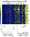
Figure 4
Figure 4. Autoantibody reactivity in Rag1S723C/S723C (mut/mut) mice after stimulation with TLR3/MDA5, TLR7/-8, and TLR9 agonists.
(A) Detection of IgG autoantibodies by protein microarray. Six- to eight-week-old mut/mut and WT/WT mice received a weekly i.p. injection of either PBS, low-dose poly(I:C), R848, or CpG. The presence of IgG antibodies against self-antigens in plasma samples was determined at day 0 and week 12 of treatment. Plasma pooled from lupus-prone NZM/MRL mice served as a positive control. Scale bars represent the MFI fold increase (blue to yellow) of autoantibody reactivity as compared with the mean + 2 SDs of the MFI in samples from WT/WT mice. (B) Frequency of autoantibodies in mut/mut mice after TLR/MDA5 stimulation. Multireactive samples were defined as containing autoantibodies against greater than or equal to 20% of 76 self-antigens. Wilcoxon test with Holm’s correction was used to compare results in WT/WT and mut/mut mice at baseline (week 0) and for each of the treatments and approached significance at week 0 (P = 0.0101). When comparing each of the 4 treatments with week 0 separately in WT/WT and in mut/mut mice, only CpG in WT/WT mice was significantly different (P = 0.0036). (C) Validation of dsDNA antibodies by ELISA. Plasma samples were diluted 200-fold. Wilcoxon test with Holm’s correction was used to compare results in WT/WT and mut/mut mice at baseline (week 0) and for each of the treatments as well as to compare each of the 4 treatments with week 0 separately in WT/WT and mut/mut mice. Results from 3 separate experiments were pooled.
Figure 3
Figure 3. Neutralizing activity of anti-cytokine antibodies.
(A) Neutralizing activity of anti–IFN-α, anti–IFN-γ, and anti–IFN-ω antibodies. Normal PBMCs were incubated in the presence of plasma from a healthy control (left panel) or from a RAG-deficient patient (right panel), and left unstimulated or stimulated with either IFN-α, IFN-γ, or IFN-ω. STAT1 phosphorylation was measured by flow cytometry. The neutralizing effect of plasma from patient CID-12 on IFN-α (red), and IFN-ω (green), but not on IFN-γ (blue), is shown as a representative example. (B) Neutralizing activity of anti–IL-12p70 antibodies. Normal lymphoblasts were incubated in the presence of plasma from a healthy control (left panel) or from RAG-deficient patient CID-1 as a representative example (right panel) and left unstimulated or stimulated with IL-12p70. STAT4 phosphorylation was measured by flow cytometry. (C) Neutralizing activity of anti–IL-22 autoantibodies. A549 adenocarcinomic human alveolar basal epithelial cells were incubated in the presence of plasma from a healthy control (left panel) or from RAG-deficient patient CID-1 as a representative example (right panel) and left unstimulated or stimulated with IL-22. STAT3 phosphorylation was measured by flow cytometry. (D) Neutralizing activity of anti–TNF-α antibodies. Jurkat 3T8 cells were incubated in the presence of plasma from a healthy control (left panel) or from RAG-deficient patient CID-12 as a representative example (right panel) and left unstimulated or stimulated with TNF-α overnight. Thy1 surface expression was measured by flow cytometry.
Figure 2
Figure 2. Anti-cytokine antibodies in RAG-deficient patients.
(A) Heatmap of autoantibody reactivity. Plasma samples from 16 healthy controls (Ctr), 14 RAG-deficient patients (OS, n = 3; LS, n = 2; CID-G/AI, n = 8; idiopathic CD4+ TCL, n = 1), and 1 patient with APS-1 were tested for anti-cytokine antibodies. The complete array is shown in the left panel, and the area with the highest reactivity is magnified on the right. Antibodies against IFN-α, IFN-ω, and IL-12 (in red) were detected with high MFI in cluster 2, including RAG-deficient patients and APS-1 patients as a positive control. (B) Elevated levels of antibodies against TPO and IFN-α were detected in RAG-deficient patients as compared with healthy controls using SAM after 10,000 permutations of the data with an FDR of less than 0.00001. (C) Multiplex bead assay for anti-cytokine antibodies. Levels of antibodies targeting IFN-α, IFN-ω, IL-12p70, IFN-γ, IFN-β, TNF-α, and IL-22 in healthy controls (n = 15) and RAG-deficient patients (n = 23), grouped by phenotype: SCID, n = 3; LS, n = 3; OS, n = 5; CID-G/AI, n = 11; and TCL, n = 1. (D) Detection of anti–IFN-α-2A, –IFN-ω, and –IL-12p70 antibodies by ELISA. Plasma samples were assayed for IgG autoantibodies at a 200-fold dilution. RAG-deficient patients included those with SCID (n = 2); LS (n = 3); OS (n = 3); CID-G/AI (n = 9); and TCL (n = 1). In both C and D, floating bars indicate the range of values for each autoantibody in healthy controls (n = 15 in C; n = 6 in D).
Figure 1
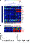
Figure 1. Autoantibodies in RAG-deficient patients as detected by protein microarray.
(A) IgG autoantibodies in 19 healthy controls (HCs) and 22 patients with RAG mutations. RAG-deficient patients were divided into 2 groups according to the severity of the clinical phenotype. Group 1 included patients with TB SCID, OS, and LS. Group 2 included patients with delayed presentation and/or a milder phenotype: CID-G/AI and TCL. MFI was normalized to that of healthy controls (mean + 2 SDs = 1), generating the RAR. Healthy controls had a lower percentage of positivity than did patients with RAG mutations (P = 0.0000008), and group 1 had a lower percentage of positivity than did group 2 (P = 0.028) as determined by Wilcoxon test with Holm’s adjustment. Samples that reacted to at least 20% of the self-antigens were defined as multireactive. (B) RAR to 11 autoantigens for which significantly higher levels of autoantibodies were found in the plasma of 5 CID-G/AI patients as compared with levels in 19 healthy controls. *P < 0.0005, **P < 0.0001, and ***P < 0.0001 by Wilcoxon test with Holm’s adjustment. Empty boxes indicate the range of RAR in healthy controls, with the bar representing the mean value. Hemo, hemocyanin; MPO, myeloperoxidase; PCNA, proliferating cell nuclear antigen; PL-12, alanyl-tRNA synthetase; PG, proteoglycan; RPLP, ribosomal phosphoprotein 0; Ro/SSA, ribonucleoprotein/Sjögren’s syndrome antigen A, 52 kDa; Tg, thyroglobulin; U1-BB’, U1 small nuclear ribonucleoprotein BB’ 9; U1-C, U1 small nuclear ribonucleoprotein C.

References

    1. Carneiro-Sampaio M, Coutinho A. Tolerance and autoimmunity: lessons at the bedside of primary immunodeficiencies. Adv Immunol. 2007;95:51–82. doi: 10.1016/S0065-2776(07)95002-6. - DOI - PubMed
    1. Liston A, Enders A, Siggs OM. Unravelling the association of partial T-cell immunodeficiency and immune dysregulation. Nat Rev Immunol. 2008;8(7):545–558. doi: 10.1038/nri2336. - DOI - PubMed
    1. Pessach I, Walter J, Notarangelo LD. Recent advances in primary immunodeficiencies: identification of novel genetic defects and unanticipated phenotypes. Pediatr Res. 2009;65(5 pt 2):3R–12R. - PubMed
    1. Schwarz K, et al. RAG mutations in human B cell-negative SCID. Science. 1996;274(5284):97–99. doi: 10.1126/science.274.5284.97. - DOI - PubMed
    1. Villa A, et al. Partial V(D)J recombination activity leads to Omenn syndrome. Cell. 1998;93(5):885–896. doi: 10.1016/S0092-8674(00)81448-8. - DOI - PubMed

Publication types

MeSH terms