. 2015 Nov;21(11):1262-1271.
doi: 10.1038/nm.3961. Epub 2015 Oct 12.

Excess TGF-β mediates muscle weakness associated with bone metastases in mice

Affiliations

Excess TGF-β mediates muscle weakness associated with bone metastases in mice

David L Waning et al. Nat Med. 2015 Nov.

Abstract

Cancer-associated muscle weakness is a poorly understood phenomenon, and there is no effective treatment. Here we find that seven different mouse models of human osteolytic bone metastases-representing breast, lung and prostate cancers, as well as multiple myeloma-exhibited impaired muscle function, implicating a role for the tumor-bone microenvironment in cancer-associated muscle weakness. We found that transforming growth factor (TGF)-β, released from the bone surface as a result of metastasis-induced bone destruction, upregulated NADPH oxidase 4 (Nox4), resulting in elevated oxidization of skeletal muscle proteins, including the ryanodine receptor and calcium (Ca(2+)) release channel (RyR1). The oxidized RyR1 channels leaked Ca(2+), resulting in lower intracellular signaling, which is required for proper muscle contraction. We found that inhibiting RyR1 leakage, TGF-β signaling, TGF-β release from bone or Nox4 activity improved muscle function in mice with MDA-MB-231 bone metastases. Humans with breast- or lung cancer-associated bone metastases also had oxidized skeletal muscle RyR1 that is not seen in normal muscle. Similarly, skeletal muscle weakness, increased Nox4 binding to RyR1 and oxidation of RyR1 were present in a mouse model of Camurati-Engelmann disease, a nonmalignant metabolic bone disorder associated with increased TGF-β activity. Thus, pathological TGF-β release from bone contributes to muscle weakness by decreasing Ca(2+)-induced muscle force production.


Figures

Figure 1
Figure 1
Skeletal muscle weakness is due to breast cancer bone metastases. (a) In vivo forelimb grip strength (n = 10) and (b) ex vivo specific force of the extensor digitorum longus (EDL) muscle in mice with MDA-MB-231 breast cancer bone metastases (n = 10). (c) Tetanic Ca2+ peak (Fluo-4) in isolated flexor digitorum brevis (FDB) muscle fibers. Representative single traces (left) and quantitation from n = 25 fibers (right). (d) Immunoblot of RyR1 oxidation (DNP) and nitrosylation (Cys NO), and RyR1-calstabin1 binding measured by co-immunoprecipitation from EDL muscle and quantitation (right). (n = 3). (e–f) RyR1 oxidation, nitrosylation and RyR1-calstabin1 binding in muscle samples from humans with (e) breast cancer bone metastases (BCa bone mets) or control samples (Ctl) and quantitation (right) (n = 4) and (f) lung cancer bone metastases (LCa bone mets) and quantitation (right) (n = 4). (g) Ex vivo EDL specific force in mice with primary MDA-MB-231 breast cancer compared to non-tumor control mice (n = 9). (h) RyR1 oxidation and nitrosylation and RyR1-calstabin1 binding in mice with primary breast cancer and quantitation (right) (n = 9). SR + H2O2 = sarcoplasmic reticulum preparations treated with hydrogen peroxide. Data are mean ± s.e.m., (a,b,g) Two-way ANOVA; (c–f) t-test; *P < 0.05, **P < 0.01, ***P < 0.001, ****P < 0.0001.
Figure 2
Figure 2
Rycal S107 improved muscle strength and lowered SR Ca2+ leak. (a) In vivo forelimb grip strength (n = 9) and (b) ex vivo specific force of the extensor digitorum longus (EDL) muscle in mice with MDA-MB-231 breast cancer bone metastases and receiving S107 treatment (n = 9). (c) Immunoblot of RyR1 oxidation (DNP) and nitrosylation (Cys NO), and RyR1-calstabin1 binding measured by co-immunoprecipitation from EDL muscle and quantitation (right) (n = 4). (d) Peak tetanic Ca2+ amplitude in mice with MDA-MB-231 bone metastases receiving S107 treatment (ratiometric imaging using Fluo-4 and Fura-red) in flexor digitorum brevis (FDB) muscle fibers (n = 36 fibers). (e) RyR1 channel open probability in muscle from mice with MDA-MB-231 bone metastases receiving S107 treatment. Representative single-channel current traces of skeletal RyR1 channels. Channel openings are shown as upward deflections. Po = open probability, To = average open time, Tc = average closed time. The activity of the channel indicated by the thick black bar is shown below on the expanded time scale. Quantification (right) of traces from n = 4 channel recordings. (f) SERCA activity from muscle of mice with bone metastases and treated with S107 (n = 4). (g) Correlation between maximum specific force (120 Hz) and osteolytic lesion area (measured in all limbs by X-ray imaging) in muscle from mice with MDA-MB-231 bone metastases receiving vehicle or S107. Data are mean ± s.e.m., (a,b) Two-way ANOVA; (c–f) One-way ANOVA with multiple comparisons; (g) Pearson's correlation. *P < 0.05, **P < 0.01, ***P < 0.001, ****P < 0.0001.
Figure 3
Figure 3
Blocking TGF-β signaling or inhibiting bone resorption lowered SMAD3 phosphorylation and improved muscle function. SMAD3 phosphorylation in (a) muscle from mice with MDA-MB-231 bone metastases and quantitation (right) (n = 4), (b) muscle samples from humans with breast cancer bone metastases (BCa bone mets) or control samples (Ctl) and quantitation (right) (n = 4) and (c) lung cancer bone metastases (LCa bone mets) and quantitation (right) (n = 4). (d) Serum TGF-β concentration in non-tumor mice, mice with MDA-MB-231 bone metastases, MDA-MB-231 primary tumor or mice with MDA-MB-231 bone metastases receiving treatment to block TGF-β signaling (SD-208) or bone resorption (zoledronic acid [ZA]) (n = 5). N.S. = not significant. (e) SMAD3 phosphorylation in mice with MDA-MB-231 bone metastases receiving either SD-208, ZA or combined therapy (n = 3). (f) In vivo forelimb grip strength (n = 12) and (g) ex vivo specific force of the extensor digitorum longus (EDL) in mice treated with SD-208, ZA or combined treatment (n = 12). (h) Immunoblot of RyR1 oxidation (DNP) and nitrosylation (Cys NO), and RyR1-calstabin1 binding measured by co-immunoprecipitation from EDL muscle and quantitation (right) (n = 3). Data are mean ± s.e.m., (a–c) t-test; (d–e,h) One-way ANOVA with multiple comparisons; (f–g) Two-way ANOVA. **P < 0.01, ***P < 0.001, ****P < 0.0001.
Figure 4
Figure 4
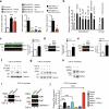
Blocking TGF-β ligand lowered SMAD3 phosphorylation and improved muscle function. (a) SMAD3 phosphorylation in muscle from mice with MDA-MB-231 bone metastases receiving 1D11 and quantitation (right) (n = 3). (b) In vivo forelimb grip strength (n = 11) and (c) ex vivo specific force of the EDL mice treated with anti-TGF-β neutralizing antibody (clone 1D11) (n = 11). (d) Immunoblot of RyR1 oxidation (DNP) and nitrosylation (Cys NO), and RyR1-calstabin1 binding measured by co-immunoprecipitation from EDL muscle and quantitation (right) (n = 3). (e) Immunoblot of RyR1 oxidation and RyR1-calstabin1 binding in C2C12 cells treated with TGF-β or TGF-β + S107. (f) Ca2+ sparks in C2C12 myotubes treated with TGF-β or TGF-β + S107 (n = 52 cells). Data are mean ± s.e.m., (a,d) t-test; (b,c) Two-way ANOVA. (f) One-way ANOVA with multiple comparisons. *P < 0.05, **P < 0.01, ****P < 0.0001.
Figure 5
Figure 5
TGF-β led to higher NADPH oxidase 4 (Nox4) expression and RyR1-Nox4 interaction. (a) Nox4 mRNA expression in tibialis anterior (TA) muscle of mice with MDA-MB-231 breast cancer bone metastases and treated with S107 (left), treated with SD-208 or zoledronic acid (center), or treated with anti-TGF-β antibody (right) (n = 3 each group). (b) Protein carbonyl concentration in muscle from mice with MDA-MB-231 bone metastases and humans with breast cancer and bone metastases (n = 2 for each group). Mean ± s.d.; Two-way ANOVA. *P<0.05 compared to Non-tumor, #P<0.05 compared to vehicle. (c–e) Nox4 co-immunoprecipitation with RyR1 in (c) muscle from mice with MDA-MB-231 bone metastases (quantitation right) (n = 3), (d) in muscle samples from humans with breast cancer and bone metastases (BCa bone mets) or control samples (Ctl) (quantitation right) (n = 4) and (e) muscle form humans with lung cancer and bone metastases (LCa bone mets) (quantitation right) (n = 4). (f) Nox4 expression in C2C12 myotubes treated with TGF-β or TGF-β + SD-208. (g) Nox4 expression in C2C12 myotubes treated with TGF-β or TGF-β + S107. (h) Nox4 co-immunoprecipitation with RyR1 in C2C12 myotubes treated with TGF-β. (i) Immunoblot in C2C12 cells with Nox4 knock-down (shNox4 or control shScr) (left), RyR1 oxidation (DNP), and RyR1-calstabin1 binding measured by co-immunoprecipitation (right). C2C12 myotubes were either untreated or treated with TGF-β. (j) Reactive oxygen species (ROS) generation in C2C12 myotubes. H2O2 = hydrogen peroxide (n = 4). Data are mean ± s.e.m., (a–b,j) One-way ANOVA with multiple comparisons; (c–e) t-test. *P < 0.05, **P < 0.01, ***P < 0.001, ****P < 0.0001.
Figure 6
Figure 6
NADPH oxidase 4 (Nox4) inhibition prevents RyR1 oxidation and improves muscle strength. (a) Immunoblot of RyR1 oxidation (DNP) and nitrosylation (Cys NO), and RyR1-calstabin1 binding measured by co-immunoprecipitation from EDL muscle and quantitation (right). (n = 4) from mice with MDA-MB-231 bone metastases and treated with Nox4 inhibitor, GKT137831. (b) Ex vivo EDL specific force in mice with MDA-MB-231 breast cancer and treated with GKT137831 (n = 8). (c) Nox4 co-immunoprecipitation with RyR1 in muscle from mice with MDA-MB-231 bone and treated with GKT137831 (quantitation right) (n = 4). (d) SMAD3 phosphorylation in muscle from mice with MDA-MB-231 bone metastases and treated with GKT137831 (quantitation right) (n = 4). (e) Model of the role of TGF-β-induced oxidation and intracellular Ca2+ leak in breast cancer-associated muscle weakness. TGF-β liberated from the bone matrix during osteolytic bone destruction drives the expression of Nox4 in muscle via SMAD2-SMAD3 signaling. Nox4 produces reactive oxygen species (ROS). Oxidation of RyR1 leads to loss of Calstabin1 binding and Ca2+ leak from the sarcoplasmic reticulum thus depleting intracellular Ca2+ stores causing decreased muscle specific force. Inhibiting TGF-β release from the bone matrix, TGF-β signaling, Nox4 activity, and RyR1-mediated SR Ca2+ leak all improved muscle strength. DHPR = dihydropyridine receptor. Data are mean ± s.e.m., (a,c–d) One-way ANOVA with multiple comparisons; (b) Two-way ANOVA. ****P < 0.0001.

References

    1. Fearon KC, Glass DJ, Guttridge DC. Cancer cachexia: mediators, signaling, and metabolic pathways. Cell Metab. 2012;16:153–166. - PubMed
    1. Gentry BA, Ferreira JA, Phillips CL, Brown M. Hindlimb skeletal muscle function in myostatin-deficient mice. Muscle Nerve. 2011;43:49–57. - PMC - PubMed
    1. Mendias CL, Marcin JE, Calerdon DR, Faulkner JA. Contractile properties of EDL and soleus muscles of myostatin-deficient mice. J. Appl. Physiol. 2006;101:898–905. - PMC - PubMed
    1. Weilbaecher KN, Guise TA, McCauley LK. Cancer to bone: a fatal attraction. Nat. Rev. Cancer. 2011;11:411–425. - PMC - PubMed
    1. Coleman RE, et al. Metastasis and bone loss: advancing treatment and prevention. Cancer Treat. Rev. 2010;36:615–620. - PMC - PubMed

Publication types

MeSH terms