. 2015 Oct 22;526(7574):591-4.
doi: 10.1038/nature15377. Epub 2015 Oct 12.

Dynamic m(6)A mRNA methylation directs translational control of heat shock response

Affiliations

Dynamic m(6)A mRNA methylation directs translational control of heat shock response

Jun Zhou et al. Nature. .

Abstract

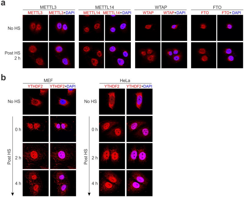
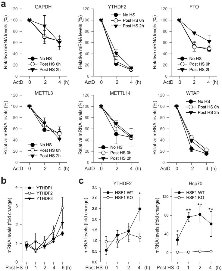
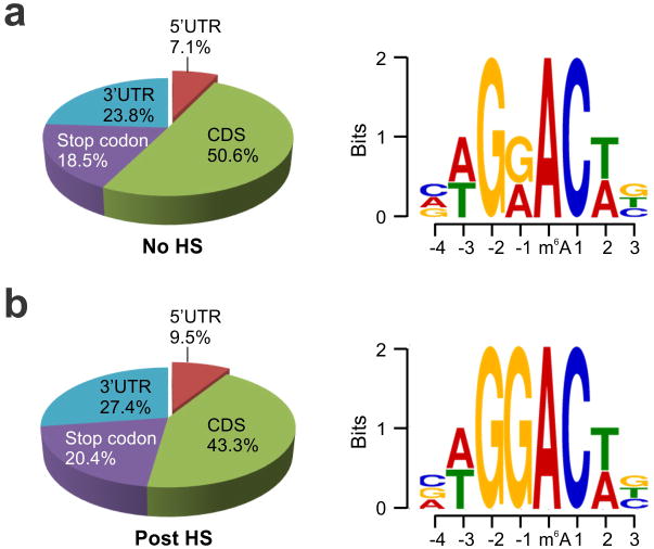
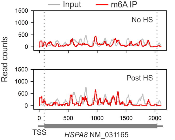
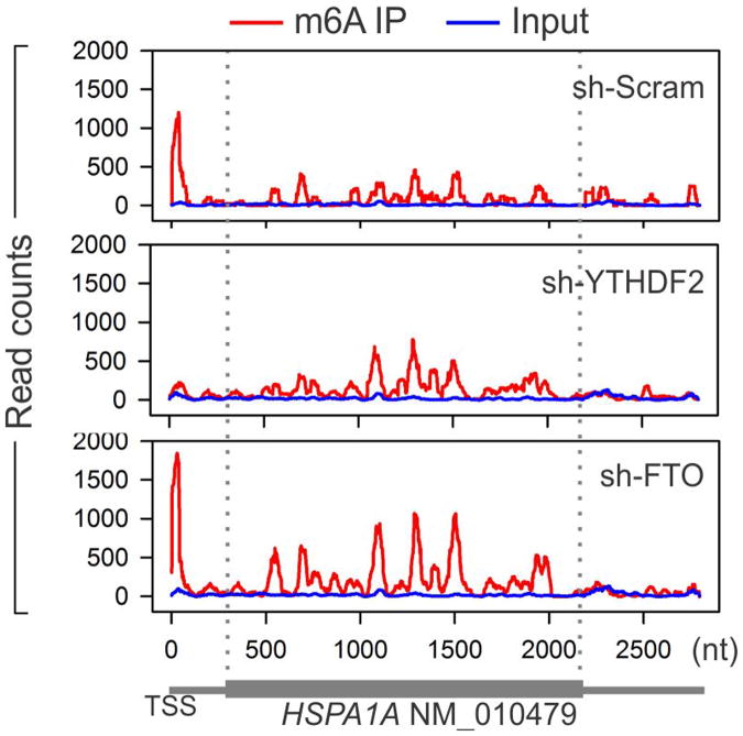
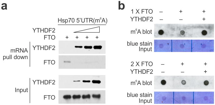
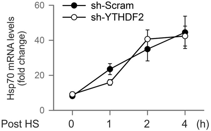
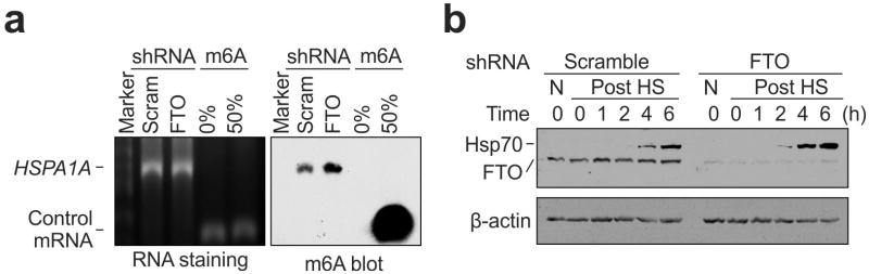
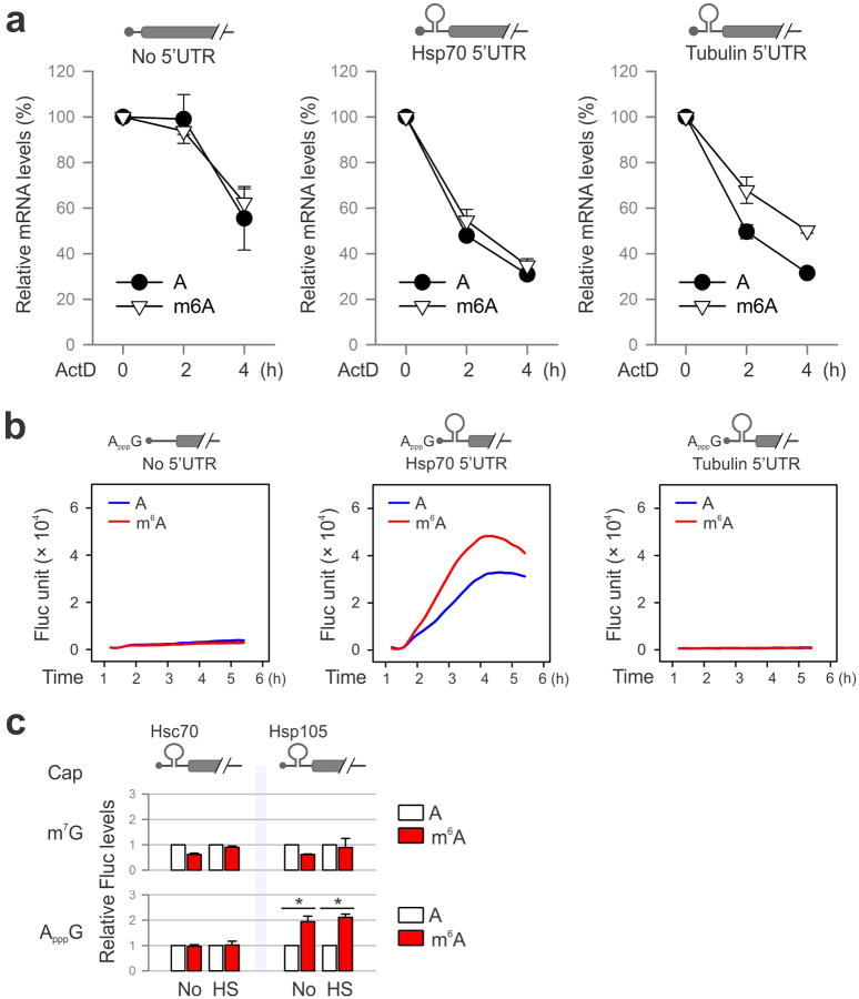
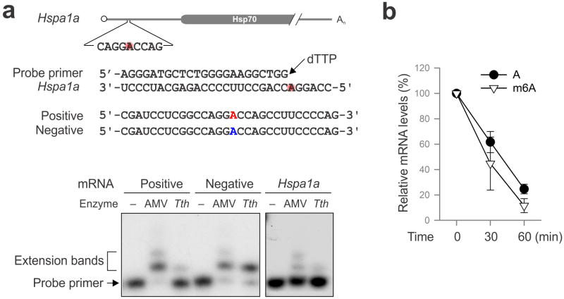
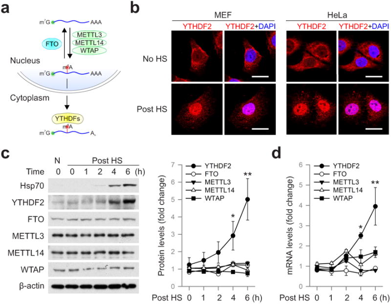
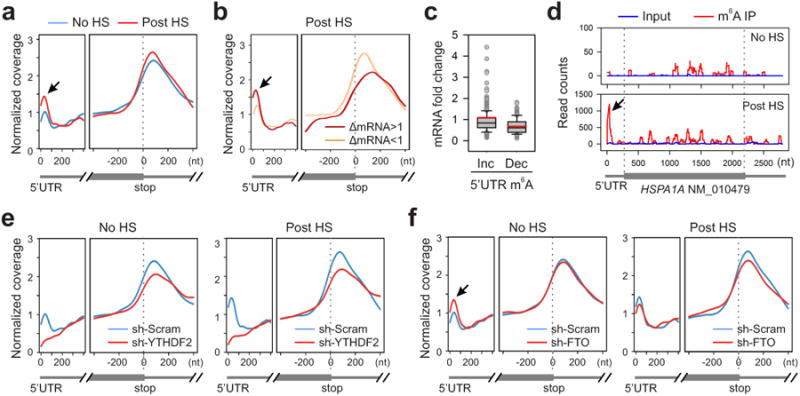
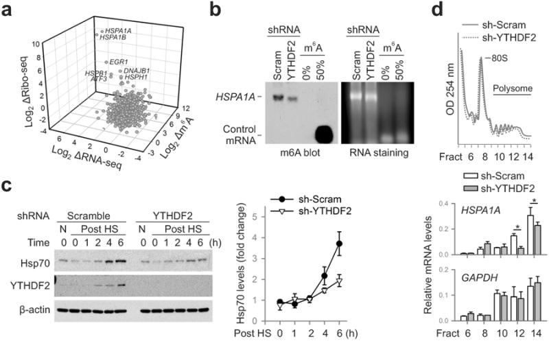
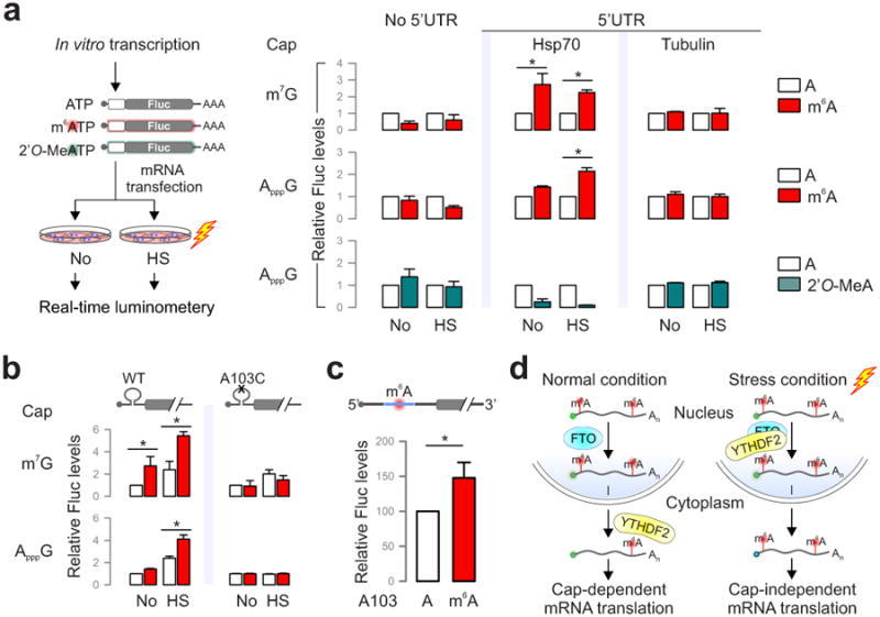
The most abundant mRNA post-transcriptional modification is N(6)-methyladenosine (m(6)A), which has broad roles in RNA biology. In mammalian cells, the asymmetric distribution of m(6)A along mRNAs results in relatively less methylation in the 5' untranslated region (5'UTR) compared to other regions. However, whether and how 5'UTR methylation is regulated is poorly understood. Despite the crucial role of the 5'UTR in translation initiation, very little is known about whether m(6)A modification influences mRNA translation. Here we show that in response to heat shock stress, certain adenosines within the 5'UTR of newly transcribed mRNAs are preferentially methylated. We find that the dynamic 5'UTR methylation is a result of stress-induced nuclear localization of YTHDF2, a well-characterized m(6)A 'reader'. Upon heat shock stress, the nuclear YTHDF2 preserves 5'UTR methylation of stress-induced transcripts by limiting the m(6)A 'eraser' FTO from demethylation. Remarkably, the increased 5'UTR methylation in the form of m(6)A promotes cap-independent translation initiation, providing a mechanism for selective mRNA translation under heat shock stress. Using Hsp70 mRNA as an example, we demonstrate that a single m(6)A modification site in the 5'UTR enables translation initiation independent of the 5' end N(7)-methylguanosine cap. The elucidation of the dynamic features of 5'UTR methylation and its critical role in cap-independent translation not only expands the breadth of physiological roles of m(6)A, but also uncovers a previously unappreciated translational control mechanism in heat shock response.


Figures

Extended Data Figure 1
Extended Data Figure 1. Subcellular localization of the m6A machinery in cells before and after heat shock stress
a, MEF cells before or 2 h after heat shock (42°C, 1 h) were immunostanied with antibodies indicated. DAPI was used for nuclear staining. b, MEF (left panel) and HeLa cells (right panel) were subject to heat shock stress (42°C, 1 h) followed by recovery at 37°C for various times. Anti-YTHDF2 immunostaining was counter stained by DAPI. Representative of at least 50 cells.
Extended Data Figure 2
Extended Data Figure 2. mRNA stability and induction in response to heat shock stress
a, Effects of heat shock stress on mRNA stability. MEF cells without heat shock stress (No HS), immediately after heat shock stress (42°C, 1 h) (Post HS 0h), or 2 h recovery at 37°C (Post HS 2h) were subject to further incubation in the presence of 5 μg/ml ActD. At the indicated times, mRNA levels were determined by qPCR. Error bars, mean ± s.e.m. n=3. b, MEF cells were collected at indicated times after heat shock stress (42°C, 1 h) followed by RNA extraction and real-time PCR. Relative levels of indicated transcripts are normalized to β-actin. Error bars, mean ± s.e.m. n=3, biological replicates. c, HSF1 WT and KO cells were subject to heat shock stress (42°C, 1 h) followed by recovery at 37°C for various times. Real-time PCR was conducted to quantify transcripts encoding Hsp70 and YTHDF2. Relative levels of transcripts are normalized to β-actin. Error bars, mean ± s.e.m. *, p < 0.05, **, p < 0.01, unpaired two-tailed t-test; n=3, biological replicates.
Extended Data Figure 3
Extended Data Figure 3. Characterization of m6A sites in MEF cells with or without heat shock stress
m6A profiling was conducted on MEF cells before (a) or 2 h after heat shock (42°C, 1 h) (b). Left, pie chart presenting fractions of m6A peaks in different transcript segments. Right, sequence logo representing the consensus motif relative to m6A.
Extended Data Figure 4
Extended Data Figure 4. m6A profiling of HSPA8 in MEF cells with or without heat shock stress
An example of constitutively expressed transcript HSPA8 in MEF cells with or without heat shock stress. Coverage of m6A IP and control reads (input) are indicated in red and gray, respectively. The transcript architecture is shown below the x-axis.
Extended Data Figure 5
Extended Data Figure 5. Dynamic m6A modification of HSPA1A by YTHDF2 and FTO
An example of stress-induced transcript HSPA1A in post-stressed MEF cells with either YTHDF2 or FTO knockdown. Coverage of m6A IP and control reads (input) are indicated in red and blue, respectively. The transcript architecture is shown below the x-axis.
Extended Data Figure 6
Extended Data Figure 6. Direct competition between YTHDF2 and FTO in m6A binding
a, Synthesized mRNA with m6A was incubated with FTO (2 μg) in the presence of increasing amount of YTHDF2 (0, 0.5, 1, 2 μg), followed by RNA pulldown and immunoblotting. b, Synthesized mRNA with m6A was incubated with FTO (1 μg in top panel and 2 μg in bottom panel) in the absence of presence of YTHDF2 (4 μg), followed by m6A dot blotting.
Extended Data Figure 7
Extended Data Figure 7. YTHDF2 knockdown does not affect Hsp70 transcription after stress
MEF cells with or without YTHDF2 knockdown were subject to heat shock stress (42°C, 1 h) followed by recovery at 37°C for various times. Real-time PCR was conducted to quantify Hsp70 mRNA levels. Error bars, mean ± s.e.m.; n=3, biological replicates.
Extended Data Figure 8
Extended Data Figure 8. FTO knockdown promotes Hsp70 synthesis
a, m6A blotting of purified HSPA1A in MEF with or without FTO knockdown. mRNAs synthesized by in vitro transcription in the absence or presence of m6A were used as control. RNA staining is shown as loading control. Representative of two biological replicates. b, MEF cells with or without FTO knockdown were collected at indicated times after heat shock stress (42°C, 1 h) followed by immunoblotting using antibodies indicated. N: no heat shock. Representative of three biological replicates.
Extended Data Figure 9
Extended Data Figure 9. m6A modification promotes cap-independent translation
a, Fluc reporter mRNAs with or without 5′UTR was synthesized in the absence or presence of m6A. The transfected MEFs were incubation in the presence of 5 μg/ml ActD. At the indicated times, mRNA levels were determined by qPCR. Error bars, mean ± s.e.m.; n=3, biological replicates. b, Fluc reporter mRNAs with or without Hsp70 5′UTR was synthesized in the absence of presence of m6A, followed by addition of a non-functional cap analog ApppG. Fluc activity in transfected MEF cells was recorded using real-time luminometry. c, Constructs expressing Fluc reporter bearing 5′UTR from Hsc70 or Hsp105 are depicted on the top. Fluc activities in transfected MEF cells were quantified and normalized to the control containing normal A. Error bars, mean ± s.e.m.; *p < 0.05, unpaired two-tailed t-test; n=3, biological replicates.
Extended Data Figure 10
Extended Data Figure 10. Site-specific detection of m6A modification on HSPA1A
a, Sequences of HSPA1A template and the DNA primer used for site-specific detection. Synthesized mRNAs containing single site m6A (red) are used as positive control. Autoradiogram shows primer extension of controls (left panel) and endogenous HSPA1A (right panel). b, Fluc mRNAs with or without m6A incorporation were incubated in the RRL at 30°C for up to 60 min. mRNA levels were determined by qPCR. Error bars, mean ± s.e.m.; n=3, biological replicates.
Figure 1
Figure 1. YTHDF2 changes cellular localization and expression levels in response to heat shock stress
a, Schematic of m6A modification machinery in mammalian cells. b, Subcellular localization of YTHDF2 in MEF and HeLa cells before or 2 h after heat shock (42°C, 1 h). Bar, 10 μm. Representative of at least 50 cells. c, Immunoblotting of MEF cells after heat shock stress (42°C, 1 h). N: no heat shock. The right panel shows the relative protein levels quantified by densitometry and normalized to β-actin. Representative of three biological replicates. d, Same samples in c were used for RNA extraction and real-time PCR. Relative levels of indicated transcripts are normalized to β-actin. Error bars, mean ± s.e.m.; *p < 0.05, **p < 0.01, unpaired two-tailed t-test; n=3, biological replicates (c and d).
Figure 2
Figure 2. Altered m6A profiles in MEF cells in response to heat shock stress
a, Metagene profiles of m6A distribution across the transcriptome of cells before or 2 h after heat shock (42°C, 1 h). Black arrow indicates the m6A peak in the 5′UTR region. b, Transcripts are stratified by different expression levels after heat shock stress, followed by metagene profiles of m6A distribution. c, A box plot depicting fold changes of mRNA levels after heat shock for transcripts showing increased or decreased m6A modification in the 5′UTR. Red line, mean value. d, An example of stress-induced transcript HSPA1A harboring m6A peaks. e, Metagene profiles of m6A distribution across the transcriptome of cells with or without YTHDF2 knockdown, before or after heat shock stress. f, Metagene profiles of m6A distribution across the transcriptome of cells with or without FTO knockdown, before or after heat shock stress.
Figure 3
Figure 3. m6A modification promotes selective translation under heat shock stress
a, A 3-D plot depicting fold changes (log2) of mRNA abundance, CDS ribosome occupancy, and 5′UTR m6A levels in MEF cells after heat shock stress. b, m6A blotting of HSPA1A purified from MEF with or without YTHDF2 knockdown. mRNAs synthesized by in vitro transcription in the absence or presence of m6A were used as control. Representative of two biological replicates. c, Immunoblotting of MEF cells with or without YTHDF2 knockdown after heat shock stress (42°C, 1 h). N: no heat shock. The right panel shows the relative protein levels quantified by densitometry and normalized to β-actin. Representative of three biological replicates. d, MEF cells with or without YTHDF2 knockdown were subject to heat shock stress followed by sucrose gradient sedimentation. Specific mRNA levels in polysome fractions were measured by qPCR. The values are first normalized to the spike in control then to the total. Error bars, mean ± s.e.m.; * p < 0.05, unpaired two-tailed t-test; n=3, biological replicates (c and d).
Figure 4
Figure 4. Selective 5′UTR m6A modification mediates cap-independent translation
a, MEF cells transfected with Fluc mRNA reporters were subject to heat shock treatment and the Fluc activity was measured by real-time luminometry. Fluc activities were quantified and normalized to the one containing normal As. b, Constructs expressing Fluc reporter with Hsp70 5′UTR or the one with A103C mutation are depicted on the top. Fluc activities in transfected MEF cells were quantified and normalized to the control containing normal A without stress. c, Fluc mRNAs bearing Hsp70 5′UTR with a single m6A site were constructed using sequential splint ligation. After in vitro translation in rabbit reticulate lysates, Fluc activities were quantified and normalized to the control lacking m6A. Error bars, mean ± s.e.m.; * p < 0.05, unpaired two-tailed t-test; n=3, biological replicates (a, b and c). d, A proposed model for dynamic m6A 5′UTR methylation in response to stress and its role in cap-independent translation. Under the normal growth condition, nuclear FTO demethylates the 5′UTR m6A from nascent transcripts and the matured transcripts are translated via a cap-dependent mechanism. Under stress conditions, nuclear localization of YTHDF2 protects the 5′UTR of stress-induced transcripts from demethylation. With enhanced 5′UTR methylation, these transcripts are selectively translated via a cap-independent mechanism.

Similar articles

Cited by

References

    1. Meyer KD, Jaffrey SR. The dynamic epitranscriptome: N6-methyladenosine and gene expression control. Nat Rev Mol Cell Biol. 2014;15:313–326. - PMC - PubMed
    1. Fu Y, Dominissini D, Rechavi G, He C. Gene expression regulation mediated through reversible m(6)A RNA methylation. Nat Rev Genet. 2014;15:293–306. - PubMed
    1. Wang X, et al. N6-methyladenosine-dependent regulation of messenger RNA stability. Nature. 2014;505:117–120. - PMC - PubMed
    1. Liu N, et al. N(6)-methyladenosine-dependent RNA structural switches regulate RNA-protein interactions. Nature. 2015;518:560–564. - PMC - PubMed
    1. Wang X, et al. N(6)-methyladenosine Modulates Messenger RNA Translation Efficiency. Cell. 2015;161:1388–1399. - PMC - PubMed

Publication types

MeSH terms

Associated data