Comparison of the two cerebral hemispheres in inhibitory processes operative during movement preparation
- PMID: 26458519
- PMCID: PMC4699690
- DOI: 10.1016/j.neuroimage.2015.10.007
Comparison of the two cerebral hemispheres in inhibitory processes operative during movement preparation
Abstract
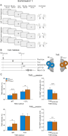
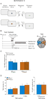
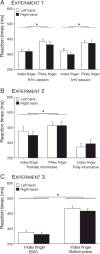
Neuroimaging and neuropsychological studies suggest that in right-handed individuals, the left hemisphere plays a dominant role in praxis, relative to the right hemisphere. However hemispheric asymmetries assessed with transcranial magnetic stimulation (TMS) has not shown consistent differences in corticospinal (CS) excitability of the two hemispheres during movements. In the current study, we systematically explored hemispheric asymmetries in inhibitory processes that are manifest during movement preparation and initiation. Single-pulse TMS was applied over the left or right primary motor cortex (M1LEFT and M1RIGHT, respectively) to elicit motor-evoked potentials (MEPs) in the contralateral hand while participants performed a two-choice reaction time task requiring a cued movement of the left or right index finger. In Experiments 1 and 2, TMS probes were obtained during a delay period following the presentation of the preparatory cue that provided partial or full information about the required response. MEPs were suppressed relative to baseline regardless of whether they were elicited in a cued or uncued hand. Importantly, the magnitude of these inhibitory changes in CS excitability was similar when TMS was applied over M1LEFT or M1RIGHT, irrespective of the amount of information carried by the preparatory cue. In Experiment 3, there was no preparatory cue and TMS was applied at various time points after the imperative signal. When CS excitability was probed in the cued effector, MEPs were initially inhibited and then rose across the reaction time interval. This function was similar for M1LEFT and M1RIGHT TMS. When CS excitability was probed in the uncued effector, MEPs remained inhibited throughout the RT interval. However, MEPs in right FDI became more inhibited during selection and initiation of a left hand movement, whereas MEPs in left FDI remained relatively invariant across RT interval for the right hand. In addition to these task-specific effects, there was a global difference in CS excitability across experiments between the two hemispheres. When the intensity of stimulation was set to 115% of the resting threshold, MEPs were larger when the TMS probe was applied over the M1LEFT than over M1RIGHT. In summary, while the latter result suggests that M1LEFT is more excitable than M1RIGHT, the recruitment of preparatory inhibitory mechanisms is similar within the two cerebral hemispheres.
Keywords: Competition; Corticospinal excitability; Decision making; Hemisphere; Inhibition; Intermanual; Response selection; Transcranial magnetic stimulation.
Copyright © 2015 Elsevier Inc. All rights reserved.
Figures


Similar articles
-
Preparatory inhibition: Impact of choice in reaction time tasks.Neuropsychologia. 2019 Jun;129:212-222. doi: 10.1016/j.neuropsychologia.2019.04.016. Epub 2019 Apr 20. Neuropsychologia. 2019. PMID: 31015024
-
Motor-evoked potentials reveal functional differences between dominant and non-dominant motor cortices during response preparation.Cortex. 2018 Jun;103:1-12. doi: 10.1016/j.cortex.2018.02.004. Epub 2018 Feb 21. Cortex. 2018. PMID: 29533856
-
Lateralized asymmetries in distribution of muscular evoked responses: An evidence of specialized motor control over an intrinsic hand muscle.Brain Res. 2018 Apr 1;1684:60-66. doi: 10.1016/j.brainres.2018.01.031. Epub 2018 Feb 3. Brain Res. 2018. PMID: 29408387
-
Interactions between cognitive and sensorimotor functions in the motor cortex: evidence from the preparatory motor sets anticipating a perturbation.Rev Neurosci. 2004;15(5):371-82. doi: 10.1515/revneuro.2004.15.5.371. Rev Neurosci. 2004. PMID: 15575492 Review.
-
Transcranial Magnetic Stimulation: Decomposing the Processes Underlying Action Preparation.Neuroscientist. 2016 Aug;22(4):392-405. doi: 10.1177/1073858415592594. Epub 2015 Jul 10. Neuroscientist. 2016. PMID: 26163320 Review.
Cited by
-
Physiological Markers of Motor Inhibition during Human Behavior.Trends Neurosci. 2017 Apr;40(4):219-236. doi: 10.1016/j.tins.2017.02.006. Epub 2017 Mar 21. Trends Neurosci. 2017. PMID: 28341235 Free PMC article. Review.
-
Lesioned hemisphere-specific phenotypes of post-stroke fatigue emerge from motor and mood characteristics in chronic stroke.Eur J Neurol. 2024 Mar;31(3):e16170. doi: 10.1111/ene.16170. Epub 2023 Dec 9. Eur J Neurol. 2024. PMID: 38069662 Free PMC article.
-
Pitfalls in Scalp High-Frequency Oscillation Detection From Long-Term EEG Monitoring.Front Neurol. 2020 Jun 2;11:432. doi: 10.3389/fneur.2020.00432. eCollection 2020. Front Neurol. 2020. PMID: 32582002 Free PMC article.
-
Considering Motor Excitability During Action Preparation in Gambling Disorder: A Transcranial Magnetic Stimulation Study.Front Psychiatry. 2020 Jun 30;11:639. doi: 10.3389/fpsyt.2020.00639. eCollection 2020. Front Psychiatry. 2020. PMID: 32695036 Free PMC article.
-
Visuomotor Correlates of Conflict Expectation in the Context of Motor Decisions.J Neurosci. 2018 Oct 31;38(44):9486-9504. doi: 10.1523/JNEUROSCI.0623-18.2018. Epub 2018 Sep 10. J Neurosci. 2018. PMID: 30201772 Free PMC article.
References
-
- Amunts K, Schlaug G, Schleicher A, Steinmetz H, Dabringhaus A, et al. Asymmetry in the human motor cortex and handedness. NeuroImage. 1996;4:216–222. - PubMed
-
- Bestmann S, Duque J. Transcranial magnetic stimulation: decomposing the processes underlying action preparation. Neuroscientist. 2015 in press. - PubMed
-
- Brainard DH. The Psychophysics Toolbox. Spat Vis. 1997;10:433–436. - PubMed
Publication types
MeSH terms
Grants and funding
LinkOut - more resources
Full Text Sources
Other Literature Sources

