Patterns of effective connectivity during memory encoding and retrieval differ between patients with mild cognitive impairment and healthy older adults
- PMID: 26458520
- PMCID: PMC5619652
- DOI: 10.1016/j.neuroimage.2015.10.002
Patterns of effective connectivity during memory encoding and retrieval differ between patients with mild cognitive impairment and healthy older adults
Abstract
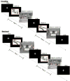
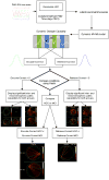
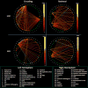
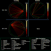
Previous research has shown that there is considerable overlap in the neural networks mediating successful memory encoding and retrieval. However, little is known about how the relevant human brain regions interact during these distinct phases of memory or how such interactions are affected by memory deficits that characterize mild cognitive impairment (MCI), a condition that often precedes dementia due to Alzheimer's disease. Here we employed multivariate Granger causality analysis using autoregressive modeling of inferred neuronal time series obtained by deconvolving the hemodynamic response function from measured blood oxygenation level-dependent (BOLD) time series data, in order to examine the effective connectivity between brain regions during successful encoding and/or retrieval of object location associations in MCI patients and comparable healthy older adults. During encoding, healthy older adults demonstrated a left hemisphere dominant pattern where the inferior frontal junction, anterior intraparietal sulcus (likely involving the parietal eye fields), and posterior cingulate cortex drove activation in most left hemisphere regions and virtually every right hemisphere region tested. These regions are part of a frontoparietal network that mediates top-down cognitive control and is implicated in successful memory formation. In contrast, in the MCI patients, the right frontal eye field drove activation in every left hemisphere region examined, suggesting reliance on more basic visual search processes. Retrieval in the healthy older adults was primarily driven by the right hippocampus with lesser contributions of the right anterior thalamic nuclei and right inferior frontal sulcus, consistent with theoretical models holding the hippocampus as critical for the successful retrieval of memories. The pattern differed in MCI patients, in whom the right inferior frontal junction and right anterior thalamus drove successful memory retrieval, reflecting the characteristic hippocampal dysfunction of these patients. These findings demonstrate that neural network interactions differ markedly between MCI patients and healthy older adults. Future efforts will investigate the impact of cognitive rehabilitation of memory on these connectivity patterns.
Published by Elsevier Inc.
Figures


References
-
- Roediger HL, Gallo DA, Geraci L. Processing approaches to cognition: The impetus from the levels-of-processing framework. Memory. 2002;10(5–6):319–332. - PubMed
-
- Spaniol J, et al. Event-related fMRI studies of episodic encoding and retrieval: meta-analyses using activation likelihood estimation. Neuropsychologia. 2009;47(8–9):1765–79. - PubMed
-
- Rugg MD, et al. Encoding-retrieval overlap in human episodic memory: A functional neuroimaging perspective. In: Sossin WS, et al., editors. Essence of Memory. 2008. pp. 339–352. - PubMed
Publication types
MeSH terms
Substances
Grants and funding
LinkOut - more resources
Full Text Sources
Other Literature Sources
Medical

