. 2015 Oct 19;212(11):1869-82.
doi: 10.1084/jem.20151403. Epub 2015 Oct 12.

Unique and redundant functions of NKp46+ ILC3s in models of intestinal inflammation

Affiliations

Unique and redundant functions of NKp46+ ILC3s in models of intestinal inflammation

Christina Song et al. J Exp Med. .

Abstract

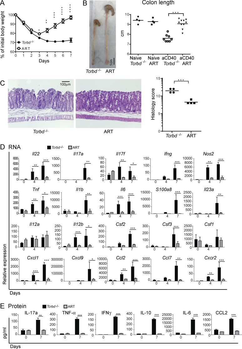
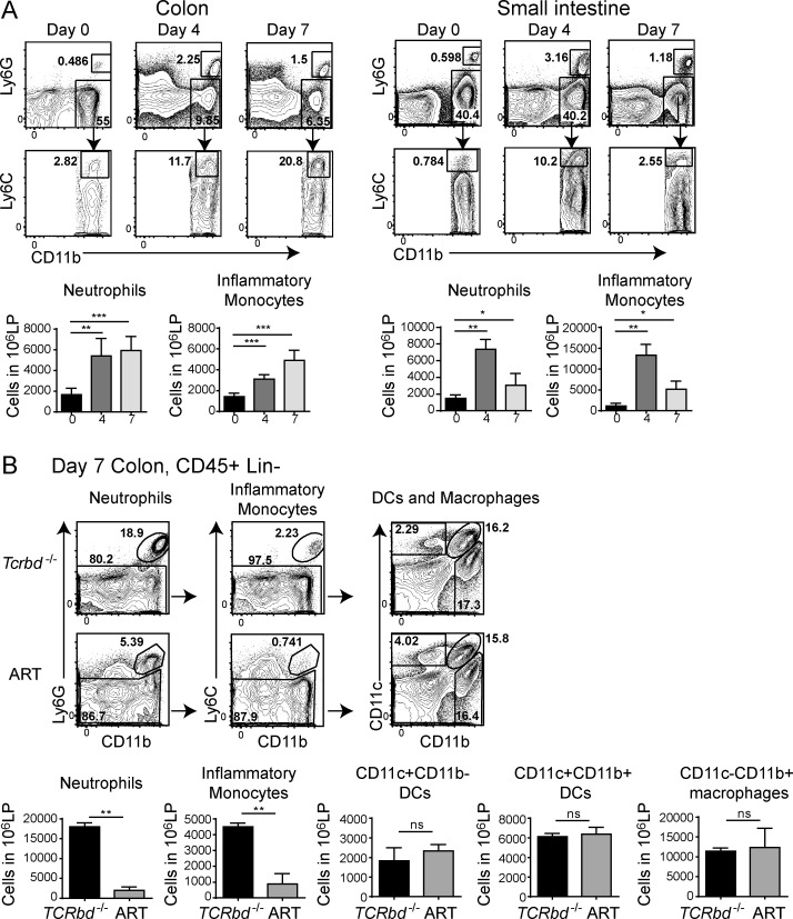
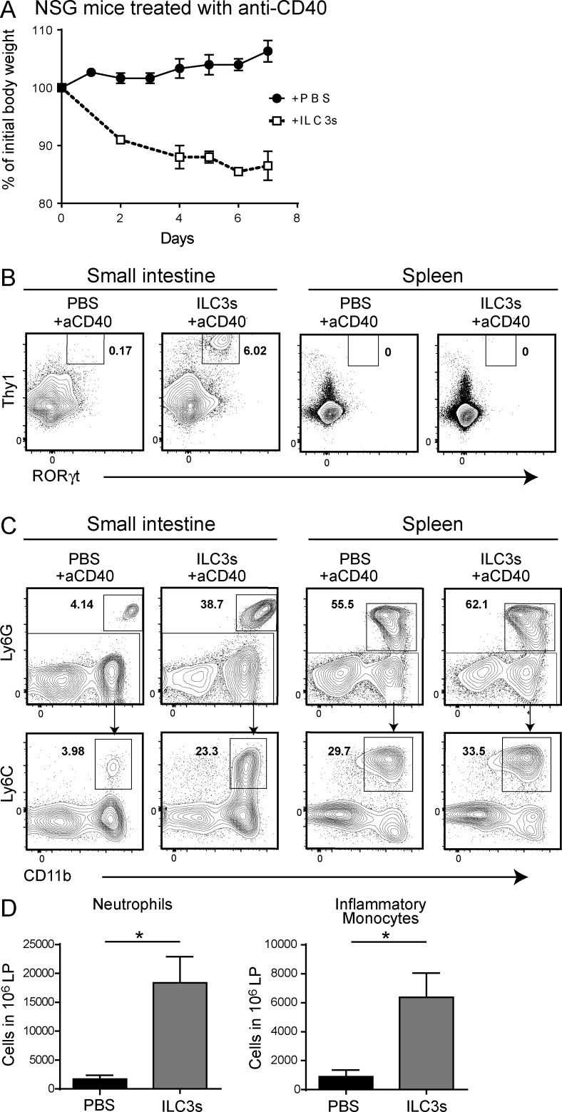
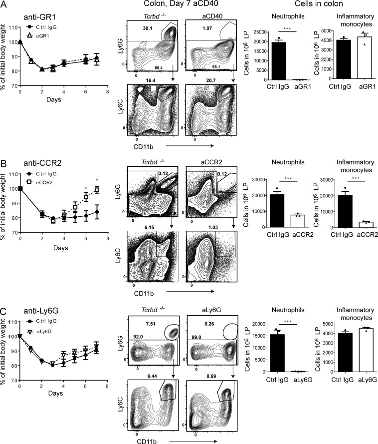
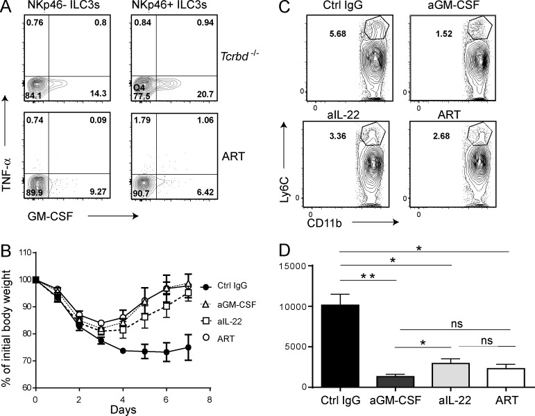
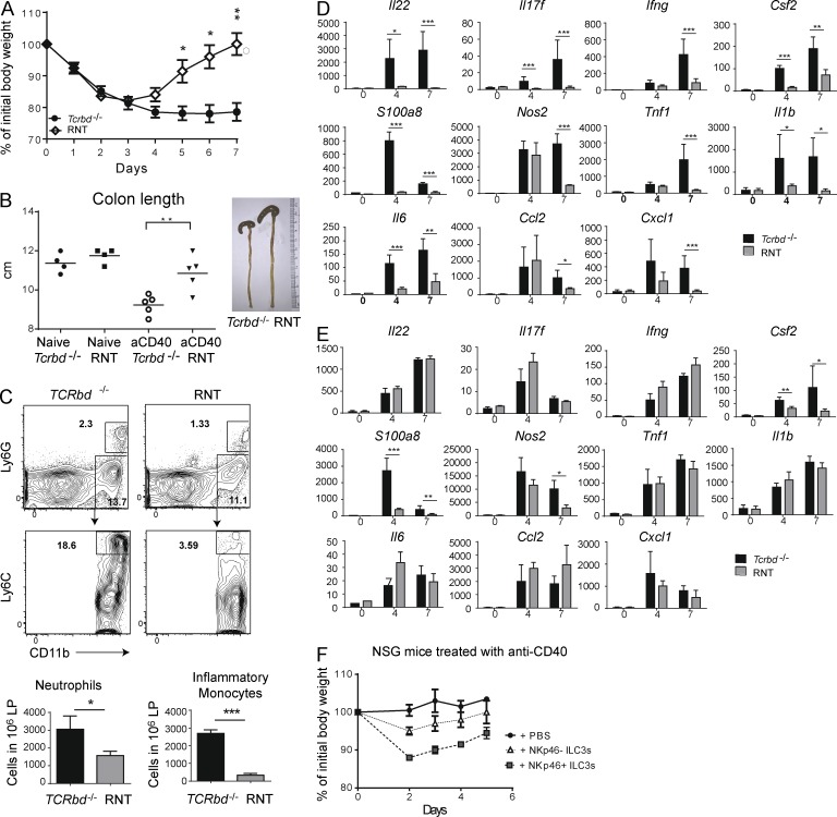
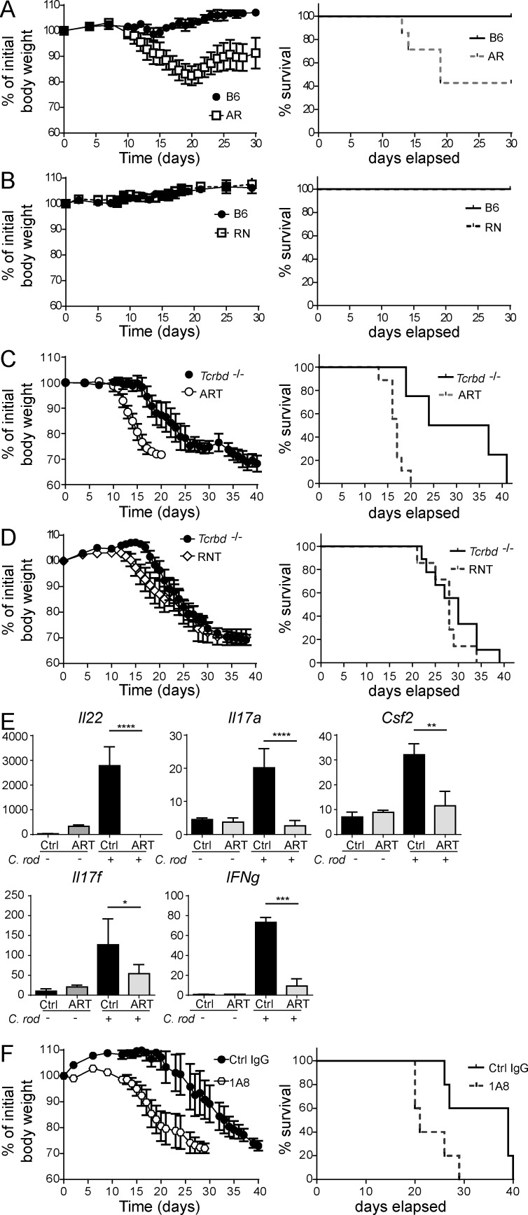
Group 3 ILCs (ILC3s) are innate sources of IL-22 and IL-17 and include lymphoid tissue-inducer (LTi)-like and NKp46(+) subsets. Both depend on RORγt and aryl hydrocarbon receptor, but NKp46(+)ILC3s also require Notch and T-bet for their development and are transcriptionally distinct. The extent to which these subsets have unique functions, especially in the context of T cell- and B cell-sufficient mice, remains largely unclear. To investigate the specific function of NKp46(+)ILC3s among other ILC3 subsets and T cells, we generated mice selectively lacking NKp46(+)ILC3s or all ILC3s and crossed them to T cell-deficient mice, thus maintaining B cells in all mice. In mice lacking T cells, NKp46(+)ILC3s were sufficient to promote inflammatory monocyte accumulation in the anti-CD40 innate colitis model through marked production of GM-CSF. In T cell-competent mice, lack of NKp46(+)ILCs had no impact on control of intestinal C. rodentium infection, whereas lack of all ILC3s partially impaired bacterial control. Thus, NKp46(+)ILC3s have a unique capacity to promote inflammation through GM-CSF-induced accumulation of inflammatory monocytes, but are superseded by LTi-like ILC3s and T cells in controlling intestinal bacterial infection.


Figures

Figure 1.
Figure 1.
ILC3 deficiency protects mice from anti-CD40–induced colitis. ART and Tcrbd−/− mice were injected with anti-CD40. (A) Mice were weighed daily, from day 0 to 7 p.i. (B) Colons were harvested and overall lengths measured 7 d p.i. Colon lengths were also assessed in Ahrf/f x Tcrbd−/− (flox control mice) and Rorγt-cre x Tcrbd−/− (Cre control mice; not depicted). A and B represent five independent experiments with 4–10 mice/group per experiment. (C) Colons were stained with hematoxylin and eosin and scored for severity of colitis on day 7, where a higher score corresponds to increased pathology. Data represent two independent experiments with four to five mice/group. (D) mRNA expression of inflammatory mediators. Colons were collected from ART and Tcrbd−/− mice at day 4 and 7 after anti-CD40 injection and the indicated transcripts were measured via quantitative RT-PCR. All gene expression values were normalized to Gapdh. (E) Protein expression of inflammatory mediators. Colonic lamina propria was processed at day 7 after anti-CD40 treatment; 106 cells were isolated and cultured for 4 h. The indicated cytokines were measured by cytometric bead array of the culture supernatants. (D and E) Data represent two independent experiments with five mice/group. *, P < 0.05; **, P < 0.01; ***, P < 0.001; ****, P < 0.0001, Student’s t test. Bars represent mean ± SEM.
Figure 2.
Figure 2.
Evaluation of intestinal myeloid populations in ART and Tcrbd−/− mice treated with anti-CD40. (A) Tcrbd−/− mice were treated with anti-CD40 and sacrificed at days 0, 4, and 7. Cells were purified from colonic and small intestinal lamina propria. Ly6G+ neutrophils and Ly6GLy6Chi inflammatory monocytes were distinguished within the population of live CD45+CD19CD11b+ cells. Representative plots (top) and neutrophil and monocyte numbers among 106 total leukocytes (bottom) are indicated. Data represent three independent experiments with two mice/time point. (B) Colonic lamina propria cells from Tcrbd−/− and ART mice were collected at day 7 after anti-CD40 treatment. Live, CD45+CD19 cells were analyzed for expression of Ly6G, Ly6C, CD11c, and CD11b. Data represent five independent experiments with two mice/group. Representative plots (top) and total numbers of neutrophils, monocytes, and DCs among 106 total leukocytes (bottom) are indicated. *, P < 0.05; **, P < 0.01; ***, P < 0.001, student’s t test. Bars represent mean ± SEM.
Figure 3.
Figure 3.
Adoptively transferred ILC3s cause severe colitis in NSG mice. 2 × 10⁴ small intestinal ILC3s (CD45+LinThy1+CD127+ cells) sort-purified from Rag1−/− mice were intravenously injected into NSG mice. (A) After 8 wk, mice were treated with anti-CD40, and body weights were measured daily, from 0 to 7 d p.i. Data represent two independent experiments with four mice/group. (B) Cells were purified from the small intestinal lamina propria. ILC3s were identified as CD45+LinThy1+RORγt+ cells. Data represent three independent experiments with two mice/group. (C) Ly6G+ neutrophils and Ly6GLy6Chi inflammatory monocytes were distinguished within the population of live CD45+CD19CD11b+ cells and (D) absolute numbers quantified. Data are representative of three experiments with two mice/group (C) or two independent experiments with five mice/group (D). *, P < 0.05, Student’s t test. Bars represent mean ± SEM.
Figure 4.
Figure 4.
Impact of inflammatory monocytes and neutrophils depletion on anti-CD40–induced colitis. Tcrbd−/− mice were treated with (A) anti-GR1 (RB6-8C5) or control IgG at days −1, 1, 3, and 5; (B) anti-CCR2 (MC-21) or control IgG at days −4, −1, 2, 3, and 4; (C) anti-Ly6G (1A8), or control IgG at days −1, 1, 3, and 5. Anti-CD40 was injected at day 0. (A–C) Numbers of intestinal neutrophils and inflammatory monocytes among 106 total leukocytes present in the colon are indicated for each antibody treatment to indicate the efficacy of depletion. *, P < 0.05; ***, P < 0.001, Student’s t test. Data are representative of three experiments (A and C) or two experiments (B) with four to five mice/group. Bars represent mean ± SEM.
Figure 5.
Figure 5.
Impact of GM-CSF and IL-22 neutralization on anti-CD40–induced colitis. (A) Representative plots show intracellular content of GM-CSF in LTi-like ILC3s (NKp46) and NKp46+ILC3s isolated from the small intestines of Tcrbd−/− and ART mice, 6 d after treatment with anti-CD40. Cells were surface stained to identify NKp46+ and NKp46 ILC3s; GM-CSF and TNF content were determined after 4-h culture in the presence of monensin. Data represent three independent experiments with two mice/group. (B and C) Tcrbd−/− mice were treated with anti–GM-CSF (MP1-22E9), anti–IL-22 (8E11), or control IgG at days 0, 2, 4, and 6. ART mice received no neutralizing antibodies. Anti-CD40 was injected into all mice at day 0. (B) Mice were weighed daily, from 0 to 7 d p.i. Data represent three independent experiments with five mice/group. (C) Representative plots of intestinal inflammatory monocytes present in the colon of antibody-treated mice. (D) Quantification of inflammatory monocytes as represented in (C). C and D represent three independent experiments with two mice/group. *, P < 0.05; **, P < 0.01, Student’s t test. Bars represent mean ± SEM.
Figure 6.
Figure 6.
Lack of NKp46+ ILC3s curbs inflammation during anti-CD40–induced colitis. RNT and Tcrbd−/− mice were injected with anti-CD40. (A) Mice were weighed daily after antibody administration. Data represent five independent experiments with five mice/group. (B) Colons from Tcrbd−/− and RNT mice were isolated 7 d after anti-CD40 injection and total lengths measured. Data represent three independent experiments with four to five mice/group. (C) Cells were purified from the small intestine LP. Ly6G+ neutrophils and Ly6GLy6Chi inflammatory monocytes were distinguished within the population of live CD45+CD19CD11b+ cells. Representative plots (left) and total neutrophil and monocyte numbers among 106 total leukocytes (right) are indicated. Data represent three independent experiments with two mice/group. Small intestines (D) and colons (E) were collected at day 7 after anti-CD40 injection, RNA was extracted and cytokine content was measured by quantitative RT-PCR. Y axis demonstrates relative transcript abundance and x axis shows day p.i. Data in D and E represent three independent experiments with five mice/group (F) NKp46+ ILC3 (CD45+Thy1+CD127+NK1.1KLRG1NKp46+) and LTi-like ILC3 (CD45+Thy1+CD127+ NK1.1KLRG1NKp46) subsets from small intestine lamina propria were sort purified from Rag1−/− mice and intravenously injected into NSG mice. After 8 wk, mice were treated with anti-CD40, and body weights were measured daily, from 0 to 7 d p.i. Data represent two independent experiments with five mice/group. *, P < 0.05; **, P < 0.01; ***, P < 0.001, Student’s t test. Bars represent mean ± SEM.
Figure 7.
Figure 7.
Contribution of ILC3s to defense against C. rodentium infection. (A–D) C. rodentium (2 × 10⁹) was administrated by oral gavage to 8–12-wk-old B6 (A and B), AR (A), RN (B), Tcrbd−/− (C and D), ART (C), and RNT (D) mice. Body weight and survival were monitored from 0 to 40 d p.i. (A–D) Data represent at least three independent experiments with 5–10 mice/group. (E) RNA was isolated from colons of Tcrbd−/− and ART mice at day 7 after injection; Il22, Il17a, Il17f, Ifng, and Csf2 were measured by RT-PCR. All gene expression values were normalized to Gapdh. Data represent three independent experiments with five mice/group. (F) Neutrophil depletion in Tcrbd−/− mice accelerates lethality to C. rodentium infection. Mice were infected and neutrophil depletion performed every 3 d by administration of 100 µg of anti-Ly6G (1A8) antibody. (E and F) Data are representative of three independent experiments with five mice/group. *, P < 0.05; **, P < 0.01; ***, P < 0.001; ****, P < 0.0001, Student’s t test.

References

    1. Artis D., and Spits H.. 2015. The biology of innate lymphoid cells. Nature. 517:293–301. 10.1038/nature14189 - DOI - PubMed
    1. Basu R., O’Quinn D.B., Silberger D.J., Schoeb T.R., Fouser L., Ouyang W., Hatton R.D., and Weaver C.T.. 2012. Th22 cells are an important source of IL-22 for host protection against enteropathogenic bacteria. Immunity. 37:1061–1075. 10.1016/j.immuni.2012.08.024 - DOI - PMC - PubMed
    1. Becher B., and Segal B.M.. 2011. T(H)17 cytokines in autoimmune neuro-inflammation. Curr. Opin. Immunol. 23:707–712. 10.1016/j.coi.2011.08.005 - DOI - PMC - PubMed
    1. Buonocore S., Ahern P.P., Uhlig H.H., Ivanov I.I., Littman D.R., Maloy K.J., and Powrie F.. 2010. Innate lymphoid cells drive interleukin-23-dependent innate intestinal pathology. Nature. 464:1371–1375. 10.1038/nature08949 - DOI - PMC - PubMed
    1. Cella M., Fuchs A., Vermi W., Facchetti F., Otero K., Lennerz J.K., Doherty J.M., Mills J.C., and Colonna M.. 2009. A human natural killer cell subset provides an innate source of IL-22 for mucosal immunity. Nature. 457:722–725. 10.1038/nature07537 - DOI - PMC - PubMed

Publication types

MeSH terms