

Innate lymphoid cells in intestinal immunity and inflammation
- PMID: 26459449
- PMCID: PMC11108440
- DOI: 10.1007/s00018-015-2055-3
Innate lymphoid cells in intestinal immunity and inflammation
Abstract
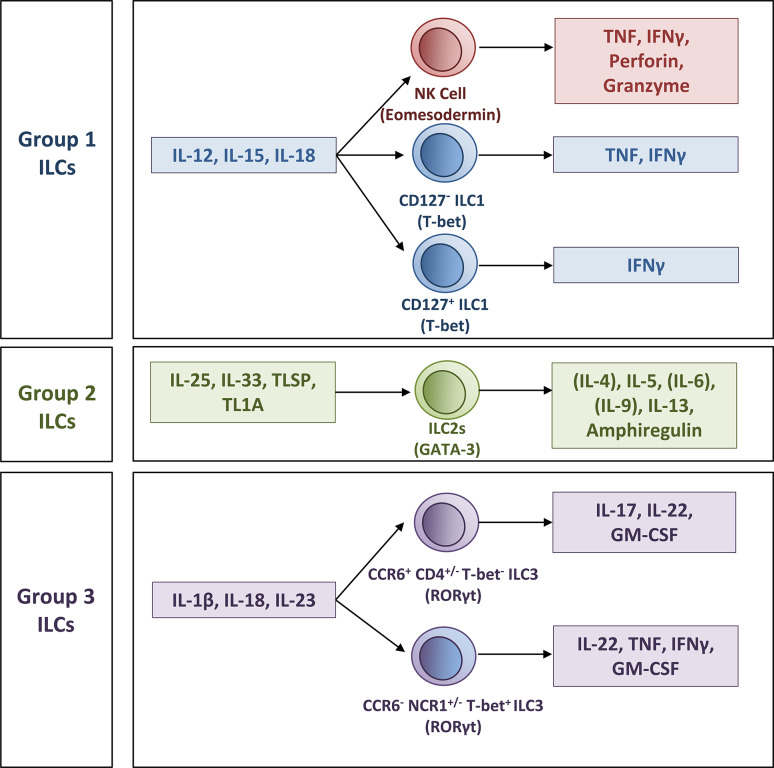
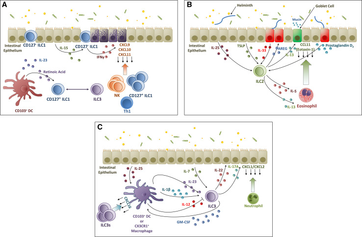
Innate lymphoid cells (ILCs) are a new and distinct family of innate immune cells that play an important role in immunity and inflammation. In this review, we focus on the role of ILCs in mucosal tissues, especially in the gut, in health and disease. ILCs support intestinal homeostasis by protecting the intestine from pathogens, contributing to the development of gut lymphoid tissue, and helping to repair injuries. By cooperating with epithelial cells and other innate and adaptive immune cells, ILCs participate in the control of pathogens and tolerance of commensal bacteria. The development and maintenance of ILCs are influenced by nutrients and metabolites sourced from diet and/or gut bacteria. ILCs have been shown to be involved in host metabolism and to participate in various diseases of the intestine including infectious and chronic inflammatory diseases, and cancer. Thus, the elucidation of ILC biology provides an exciting potential for development of novel therapeutic means to modulate immune responses in various disease settings.
Keywords: Commensals; Diet; Homeostasis; Metabolism; Nutrition.
Figures

Similar articles
-
Aryl hydrocarbon receptor promotes RORγt⁺ group 3 ILCs and controls intestinal immunity and inflammation.Semin Immunopathol. 2013 Nov;35(6):657-70. doi: 10.1007/s00281-013-0393-5. Epub 2013 Aug 23. Semin Immunopathol. 2013. PMID: 23975386 Free PMC article. Review.
-
Innate Lymphoid Cells in Intestinal Homeostasis and Inflammatory Bowel Disease.Int J Mol Sci. 2021 Jul 16;22(14):7618. doi: 10.3390/ijms22147618. Int J Mol Sci. 2021. PMID: 34299236 Free PMC article. Review.
-
Innate lymphoid cells regulate CD4+ T-cell responses to intestinal commensal bacteria.Nature. 2013 Jun 6;498(7452):113-7. doi: 10.1038/nature12240. Epub 2013 May 22. Nature. 2013. PMID: 23698371 Free PMC article.
-
The interaction of intestinal microbiota and innate lymphoid cells in health and disease throughout life.Immunology. 2020 Jan;159(1):39-51. doi: 10.1111/imm.13138. Epub 2019 Nov 27. Immunology. 2020. PMID: 31777064 Free PMC article. Review.
-
Group 3 ILCs: Peacekeepers or Troublemakers? What's Your Gut Telling You?!Front Immunol. 2019 Apr 5;10:676. doi: 10.3389/fimmu.2019.00676. eCollection 2019. Front Immunol. 2019. PMID: 31024537 Free PMC article. Review.
Cited by
-
Innate Lymphoid Cells: A Link between the Nervous System and Microbiota in Intestinal Networks.Mediators Inflamm. 2019 Jan 20;2019:1978094. doi: 10.1155/2019/1978094. eCollection 2019. Mediators Inflamm. 2019. PMID: 30804706 Free PMC article. Review.
-
Close Encounters of Lymphoid Cells and Bacteria.Front Immunol. 2016 Oct 7;7:405. doi: 10.3389/fimmu.2016.00405. eCollection 2016. Front Immunol. 2016. PMID: 27774092 Free PMC article. Review.
-
Gut Bacteria Induce Granzyme B Expression in Human Colonic ILC3s In Vitro in an IL-15-Dependent Manner.J Immunol. 2021 Jun 15;206(12):3043-3052. doi: 10.4049/jimmunol.2000239. Epub 2021 Jun 11. J Immunol. 2021. PMID: 34117105 Free PMC article.
-
The intestinal barrier in multiple sclerosis: implications for pathophysiology and therapeutics.Brain. 2018 Jul 1;141(7):1900-1916. doi: 10.1093/brain/awy131. Brain. 2018. PMID: 29860380 Free PMC article. Review.
-
Regulation of Innate Lymphoid Cells by Aryl Hydrocarbon Receptor.Front Immunol. 2018 Jan 5;8:1909. doi: 10.3389/fimmu.2017.01909. eCollection 2017. Front Immunol. 2018. PMID: 29354125 Free PMC article. Review.
References
Publication types
MeSH terms
Grants and funding
LinkOut - more resources
Full Text Sources
