. 2015 Oct 12;211(1):53-61.
doi: 10.1083/jcb.201504042.

Caveolae protect endothelial cells from membrane rupture during increased cardiac output

Affiliations

Caveolae protect endothelial cells from membrane rupture during increased cardiac output

Jade P X Cheng et al. J Cell Biol. .

Abstract

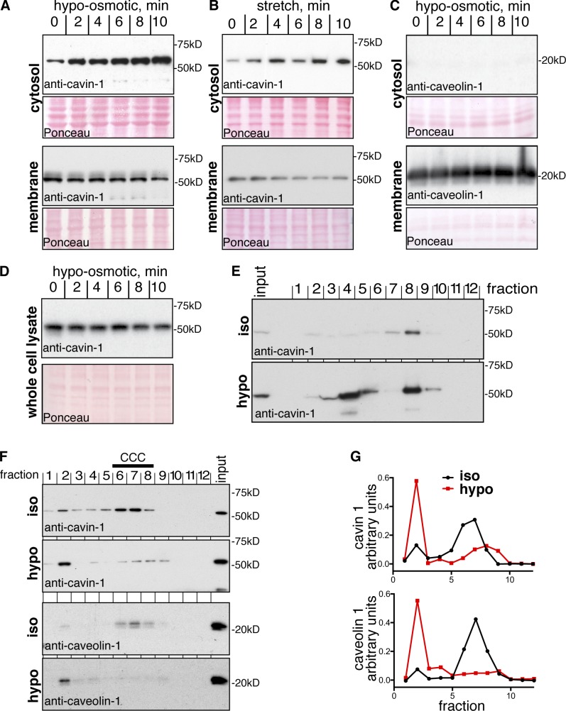
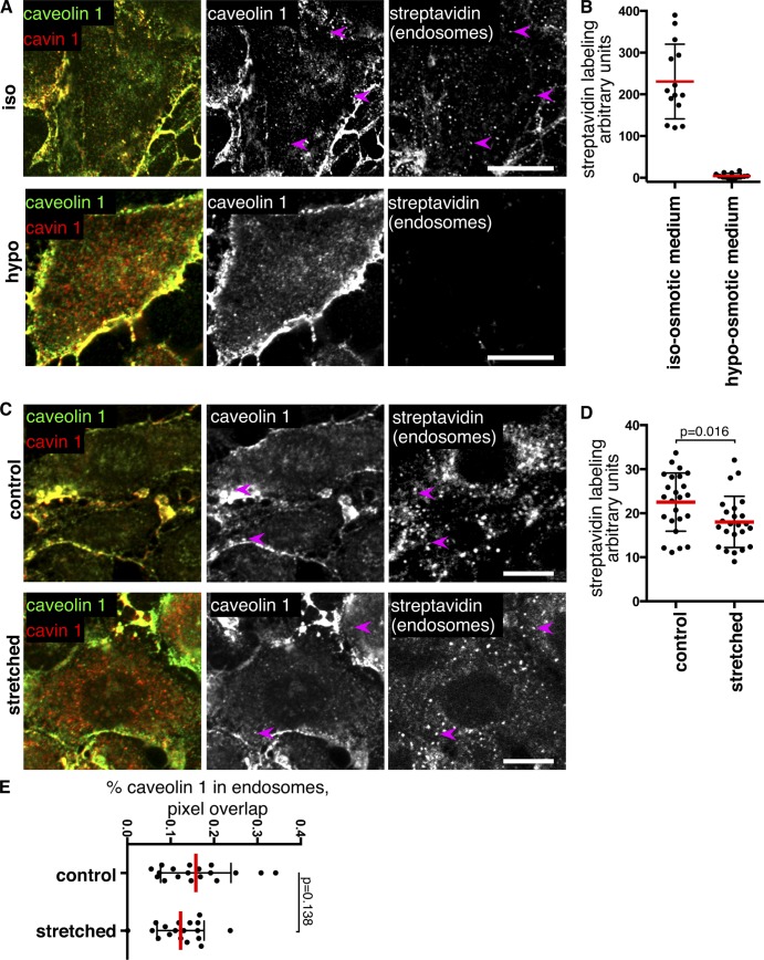
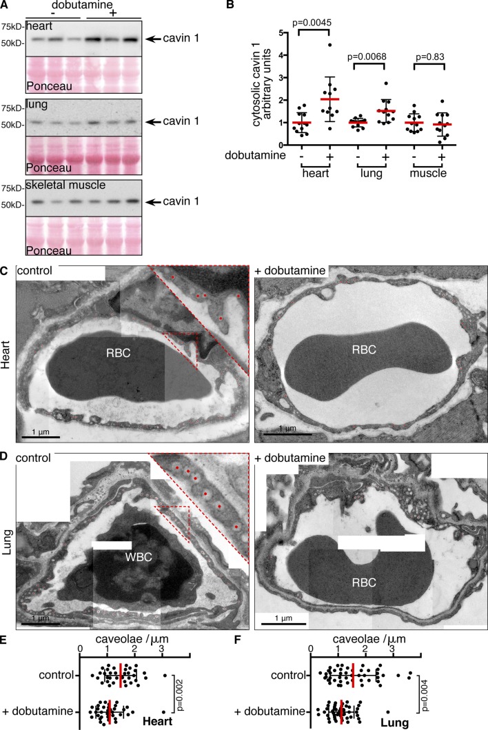
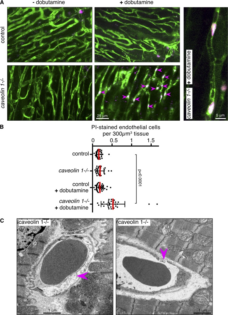
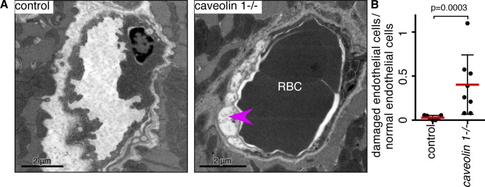
Caveolae are strikingly abundant in endothelial cells, yet the physiological functions of caveolae in endothelium and other tissues remain incompletely understood. Previous studies suggest a mechanoprotective role, but whether this is relevant under the mechanical forces experienced by endothelial cells in vivo is unclear. In this study we have sought to determine whether endothelial caveolae disassemble under increased hemodynamic forces, and whether caveolae help prevent acute rupture of the plasma membrane under these conditions. Experiments in cultured cells established biochemical assays for disassembly of caveolar protein complexes, and assays for acute loss of plasma membrane integrity. In vivo, we demonstrate that caveolae in endothelial cells of the lung and cardiac muscle disassemble in response to acute increases in cardiac output. Electron microscopy and two-photon imaging reveal that the plasma membrane of microvascular endothelial cells in caveolin 1(-/-) mice is much more susceptible to acute rupture when cardiac output is increased. These data imply that mechanoprotection through disassembly of caveolae is important for endothelial function in vivo.


Figures

Figure 1.
Figure 1.
Loss of plasma membrane caveolae under mechanical force is accompanied by disassembly of caveolar protein complexes. (A) Immunoblot analyses of cavin 1 in cytosolic and membrane fractions from cells lysed after different times in hypo-osmotic medium. Ponceau staining of the relevant areas of the blot membrane is included to demonstrate equal loading. (B) Immunoblot analyses of cavin 1 in cytosolic and membrane fractions from cells lysed after different times of mechanical stretching, the deformable substrate on which the cells were grown was stretched by 20%, repeated at 1 Hz. (C) Immunoblot analyses of caveolin 1 in cytosolic and membrane fractions from cells lysed after different times in hypo-osmotic medium. Ponceau staining of the relevant areas of the blot membrane is included to demonstrate equal loading. (D) Immunoblot analyses of cavin 1 whole cell lysates after different times in hypo-osmotic medium. (E) Cytosolic fractions from cells incubated for 10 min in isotonic (iso) or hypotonic (hypo) buffer were applied to a 10–40% sucrose velocity gradient, and analyzed by Western blotting of fractions 1–12. (F) Cross-linked and detergent-solubilized (1% Triton X-100/1% octyl-glucoside) cell extracts were fractionated by velocity centrifugation (10–40% sucrose), followed by Western blotting of fractions 1–12. Gradients were prepared from cells incubated in isotonic or hypotonic buffer for 10 min. The high molecular weight peak of caveolin 1 and cavin 1 corresponding to the caveolar coat complex (CCC) in isotonic medium is indicated. (G) Quantification of the distribution of cavin 1 and caveolin 1 in velocity gradients as shown in F. Protein amounts normalized such that area under each curve equal to 1 was determined by densitometry of Western blots.
Figure 2.
Figure 2.
Mechanical force does not induce endocytosis of caveolae. (A) Surface biotinylation using reducible sulfo-NHS-SS-biotin was followed by incubation for 10 min in isotonic or hypotonic medium at 37°C as shown. After MESNa treatment to remove noninternalized biotin, cells were fixed and labeled with antibodies against caveolin 1 and cavin 1, and with fluorescent streptavidin. Arrows highlight limited presence of caveolin 1 in endocytic structures. Note that the streptavidin channel is omitted from the red/green overlay images. Bars, 10 µm. (B) Quantification of biotin uptake after surface labeling with sulfo-NHS-SS-biotin and MESNa treatment. Mean fluorescence intensity of multiple cell regions after subtraction of background signal is shown. (C) Cells were labeled with sulfo-NHS-SS-biotin, followed by incubation for 10 min at 37°C, whereas the deformable substrate on which the cells were grown was stretched by 20%, repeated at 1 Hz. After MESNa treatment to remove noninternalized biotin, cells were fixed and labeled with antibodies against caveolin 1 and cavin 1, and with fluorescent streptavidin. Bars, 10 µm. (D) Quantification of biotin uptake after surface labeling with sulfo-NHS-SS-biotin and MESNa treatment. Mean fluorescence intensity of multiple cell regions after subtraction of background signal is shown. (E) Quantification of the proportion of caveolin 1 present in biotin-positive (i.e., endosomal) pixels after incubation as in C.
Figure 3.
Figure 3.
Increased cardiac output causes caveolar disassembly in vivo. (A) Immunoblot analyses of cavin-1 in cytosolic fractions from tissues snap frozen after 1 min of treatment of mice with dobutamine. Samples from three separate animals are shown for each tissue/treatment. Ponceau stain of the relevant Western blots is also included, to confirm equal loading. (B) Quantification of immunoblots of cytosolic cavin 1 as in A. Densitometry data in arbitrary units were normalized such that the mean of the control (i.e., without dobutamine) for each tissue is equal to 1. Each point represents one animal; mice were 20–25 wk old. (C) Multiple electron micrographs to represent endothelium of microvessels from the heart of control and dobutamine-treated mice. Red dots indicate structures counted as caveolae open to the luminal side of the endothelial cell. Dobutamine treatment was for 1 min before rapid dissection and tissue fixation. Zoomed-in regions are shown as outlined. (D) Electron micrographs of microvessels from the lung of control and dobutamine-treated mice, as in C. (E) Quantification of caveolae in microvascular endothelium of the heart. Dobutamine treatment was for 1 min. Each point represents one complete reconstruction of the endothelium from multiple micrographs as in C; data are from three control and three treated mice. (F) As in E, but tissue is lung. RBC and WBC denote red and white blood cells, respectively.
Figure 4.
Figure 4.
Endothelial caveolae protect cells from membrane damage during acute increases in cardiac output in vivo. (A) Maximum intensity projections of z stacks of two-photon images (∼20 µm deep) of heart tissue from control and caveolin 1−/− mice injected with FITC-albumin (green, capillaries) and propidium iodide (PI; magenta, nuclei), with and without injection of dobutamine. Arrows show PI-positive nuclei within capillaries. The zoomed-in view in the right-hand panel confirms that PI-positive nuclei are in capillaries. (B) Quantification of PI-positive nuclei within capillaries. Each point represents one image stack, greater than five animals were analyzed per condition. (C) Abnormal endothelial morphology in cardiac muscle of caveolin 1−/− mice immediately after dobutamine treatment. Examples of ruptured cells and of abnormal gaps between cells are arrowed.
Figure 5.
Figure 5.
Endothelial caveolae protect cells from membrane damage during hypoxia in vivo. (A) Electron micrographs of microvessels from the right ventricle of heart from mice exposed to hypoxia for 3 wk. Magenta arrowhead highlights an abnormal endothelial cell with loss of cytoplasmic staining. (B) Quantification of endothelial cells with abnormal morphology after 3 wk hypoxia. Each dot represents one animal, >20 vessels were examined for each animal.

References

    1. Albinsson S., Nordström I., Swärd K., and Hellstrand P.. 2008. Differential dependence of stretch and shear stress signaling on caveolin-1 in the vascular wall. Am. J. Physiol. Cell Physiol. 294:C271–C279. 10.1152/ajpcell.00297.2007 - DOI - PubMed
    1. Andrews N.W., Almeida P.E., and Corrotte M.. 2014. Damage control: cellular mechanisms of plasma membrane repair. Trends Cell Biol. 24:734–742. 10.1016/j.tcb.2014.07.008 - DOI - PMC - PubMed
    1. Ardissone A., Bragato C., Caffi L., Blasevich F., Maestrini S., Bianchi M.L., Morandi L., Moroni I., and Mora M.. 2013. Novel PTRF mutation in a child with mild myopathy and very mild congenital lipodystrophy. BMC Med. Genet. 14:89. 10.1186/1471-2350-14-89 - DOI - PMC - PubMed
    1. Austin E.D., Ma L., LeDuc C., Berman Rosenzweig E., Borczuk A., Phillips J.A. III, Palomero T., Sumazin P., Kim H.R., Talati M.H., et al. . 2012. Whole exome sequencing to identify a novel gene (caveolin-1) associated with human pulmonary arterial hypertension. Circ Cardiovasc Genet. 5:336–343. 10.1161/CIRCGENETICS.111.961888 - DOI - PMC - PubMed
    1. Bitsikas V., Corrêa I.R. Jr., and Nichols B.J.. 2014. Clathrin-independent pathways do not contribute significantly to endocytic flux. eLife. 3:e03970. - PMC - PubMed

Publication types

LinkOut - more resources