. 2015 Nov 1;212(9):1509-20.
doi: 10.1093/infdis/jiv221.

Ifit1 Protects Against Lipopolysaccharide and D-galactosamine-Induced Fatal Hepatitis by Inhibiting Activation of the JNK Pathway

Affiliations

Ifit1 Protects Against Lipopolysaccharide and D-galactosamine-Induced Fatal Hepatitis by Inhibiting Activation of the JNK Pathway

Antao Chang et al. J Infect Dis. .

Abstract

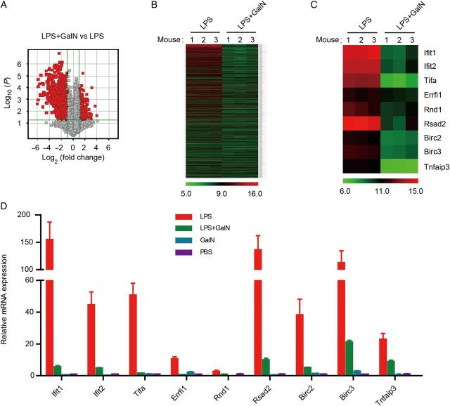
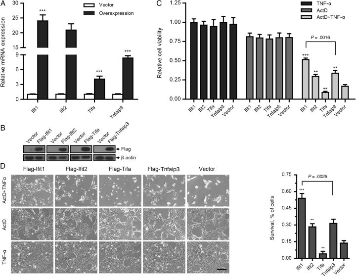
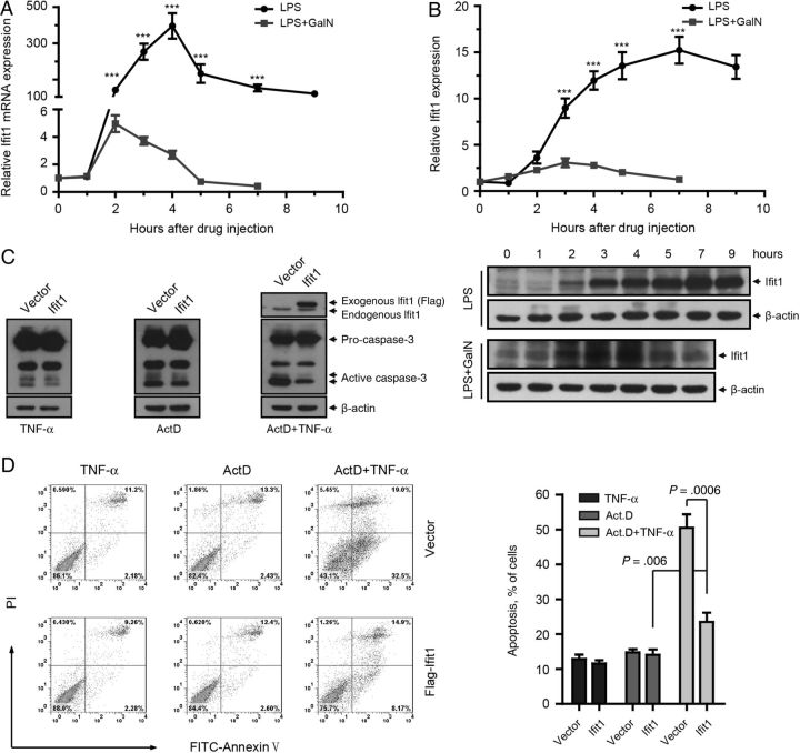
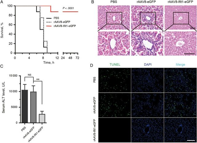
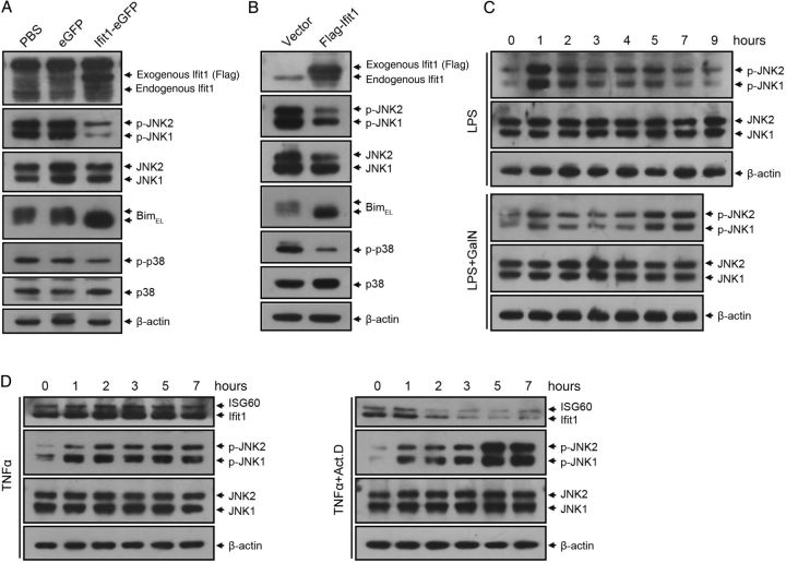
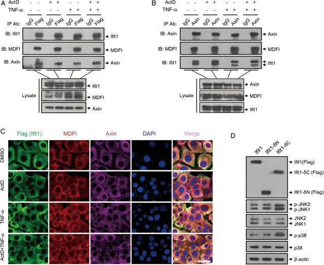
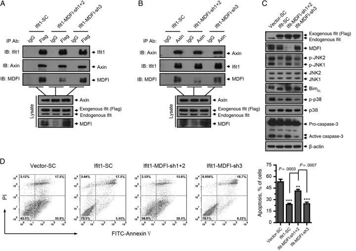
Treatment of mice with lipopolysaccharide (LPS) and the liver-specific transcriptional inhibitor D-(+)-galactosamine (GalN) induces fatal hepatitis, which is mediated by tumor necrosis factor α (TNF-α) and characterized by massive hepatic apoptosis. Previous studies suggest that GalN increases the sensitivity to LPS/TNF-α, probably by blocking the transcription of protective factors, but the identity of most of these factors is still unclear. Here, we report that Ifit1 protects against LPS/GalN-induced fatal hepatitis. Forced expression of Ifit1 in hepatocytes significantly diminished TNF-α-mediated apoptosis. Moreover, targeted expression of Ifit1 in the liver by recombinant adeno-associated virus serotype 8 protected mice from LPS/GalN-induced lethal hepatitis, which was associated with the inhibition of TNF-α-mediated activation of the c-Jun N-terminal kinase (JNK)-Bim cascade. Furthermore, Ifit1 bound to a scaffolding protein Axin and inhibited its function to mediate JNK activation. Together, our data demonstrate that Ifit1 is a novel protective factor that inhibits LPS/GalN-induced (TNF-α-mediated) fatal hepatitis, suggesting that Ifit1 is a potential therapeutic target for treatment of inflammatory liver diseases.

Keywords: Ifit1; JNK; LPS; TNF-α; fatal hepatitis.


Figures

Figure 1.
Figure 1.
Gene array analysis for protective factors that restrict lipopolysaccharide (LPS)–induced liver injury. Mice were treated with 0.01 µg of LPS alone or together with 20 mg of D-(+)-galactosamine (GalN) for 2.5 hours. Liver tissue specimens were isolated for gene array analysis. A, Volcano plots for visualizing differential gene expression between LPS and LPS/GalN treatments. B and C, Heat map depicting the messenger RNA (mRNA) levels of differentially expressed genes that were downregulated in the model. D, Real-time quantitative polymerase chain reaction analysis of the mRNA levels of genes of interest in mouse livers 2.5 hours after treatment (n = 3). Abbreviation: PBS, phosphate buffered saline.
Figure 2.
Figure 2.
Expression of some genes of interest (GOIs) reduces tumor necrosis factor α (TNF-α)–induced cell death. BNL CL.2 cells expressing GOIs or empty vector were treated with 0.01 µg/mL TNF-α and 1 µg/mL actinomycin D (ActD) separately or simultaneously for 7 hours. A and B, Real-time quantitative polymerase chain reaction and immunoblotting analyses of the overexpression efficiency of GOIs. C, Cell viability assay, based on quantitation of adenosine triphosphate (n = 3). Cell viability is relative to the same type of cells treated with dimethyl sulfoxide. D, Cell counting analysis of survival among cells (n = 3). The survival percentage is relative to the group with that received TNF-α treatment. All data are presented as mean values ± SD. Scale bar represents 100 µm. **P < .01 and ***P < .001, by the 2-tailed t test, for comparison to groups that received empty vector. Abbreviation: mRNA, messenger RNA.
Figure 3.
Figure 3.
Expression of Ifit1 protects against tumor necrosis factor α (TNF-α)–mediated apoptosis in vitro. A and B, Mice were treated with 0.01 µg of lipopolysaccharide (LPS) alone or together with 20 mg D-(+)-galactosamine (GalN). Livers were isolated at indicated times after injection. A, Real-time quantitative polymerase chain reaction analysis of Ifit1 in mouse livers (n = 3). Expression levels are normalized to levels in untreated samples. B, Immunoblotting for Ifit1 in mice liver. Curves represent quantification of the immunoblotting results by ImageJ software, and expression levels are normalized to that in lane 1 (n = 3). Data are presented as mean values ± SD. ***P < .001, by the 2-tailed t test, for comparisons to LPS/GalN groups. C and D, BNL CL.2 cells expressing Ifit1 or empty vector were treated with 0.01 µg/mL TNF-α and 1 µg/mL actinomycin D (ActD) simultaneously or separately for 7 hours. C, Immunoblotting analysis of caspase-3 activation. D, Flow cytometry analysis of apoptosis. Comparisons were made using the 2-tailed t test. Abbreviation: mRNA, messenger RNA.
Figure 4.
Figure 4.
Gene transfer of Ifit1 protects mice against lipopolysaccharide (LPS)/D-(+)-galactosamine (GalN)–induced lethal hepatitis. Mice were injected intravenously with recombinant adeno-associated virus serotype 8 (rAAV8) encoding Ifit1-eGFP or eGFP or with saline and challenged 4 days later with 0.01 µg of LPS plus 20 mg of GalN. Livers and serum were harvested 7 hours after LPS/GalN challenge. A, Survival analysis of the mice injected with indicated rAAV8. B, Histological examination of hematoxylin-eosin–stained liver sections. C, Serum alanine aminotransferase (ALT) levels (n = 3). Data are presented as mean values ± SD. Comparisons were made using the 2-tailed t test. D, Terminal deoxynucleotidyl transferase dUTP nick end labeling (TUNEL) staining of liver sections for apoptosis detection. All scale bars represent 100 µm. **P < .01. Abbreviations: NS, not significant; PBS, phosphate-buffered saline.
Figure 5.
Figure 5.
Ifit1 inhibits tumor necrosis factor α (TNF-α)–mediated activation of JNK and p38. A, Mice were injected with recombinant adeno-associated virus serotype 8 (rAAV8) or saline and challenged 4 days later with 0.01 µg of lipopolysaccharide (LPS) plus 20 mg of D-(+)-galactosamine (GalN). Liver extracts were harvested 7 hours after LPS/GalN challenge and probed by immunoblotting with antibodies against Ifit1, JNK, Bim, p38, and β-actin (loading control). B, BNL CL.2 cells expressing Ifit1 or empty vector were cotreated with 0.01 µg/mL TNF-α and 1 µg/mL actinomycin D (ActD) for 7 hours. Cell lysates were probed by immunoblotting for Ifit1, JNK, Bim, p38, and β-actin. C, Immunoblotting for JNK in mice treated with 0.01 µg of LPS alone or together with 20 mg of GalN. D, Immunoblotting for JNK in BNL CL.2 cells treated with 0.01 µg/mL TNF-α alone or together with 1 µg/mL ActD. Abbreviations: JNK, c-Jun N-terminal kinase; PBS, phosphate-buffered saline.
Figure 6.
Figure 6.
Ifit1 inhibits tumor necrosis factor α (TNF-α)–induced apoptosis by binding to Axin. A–C, BNL CL.2 cells expressing Ifit1 were or were not treated with TNF-α and actinomycin D (ActD) for 5 hours. A, Coimmunoprecipitation with an anti-Flag antibody or control immunoglobulin G (IgG), followed by immunoblotting. B, Coimmunoprecipitation with an anti-Axin antibody or control IgG, followed by immunoblotting. C, Immunofluorescence staining for detecting the localization of Ifit1, MDFI, and Axin. D, BNL CL.2 cells expressing Ifit1-δN or Ifit1-δC were cotreated with TNF-α and ActD for 5 hours. Cell lysates were probed by immunoblotting for Ifit1, JNK, p38, and β-actin. Scale bar represents 30 µm. Abbreviations: JNK, c-Jun N-terminal kinase; DMSO, dimethyl sulfoxide.
Figure 7.
Figure 7.
MDFI is not required for the physical interaction between Ifit1 and axin, but it is necessary for Ifit1-mediated inhibition of axin. BNL CL.2 cells expressing Ifit1 or empty vector were infected with lentivirus encoding small hairpin RNA for MDFI or β-galactosidase from Escherichia coli (SC) and then were cotreated with tumor necrosis factor α (TNF-α) and actinomycin D (ActD) for 5–7 hours. A, Coimmunoprecipitation with an anti-Flag antibody or control immunoglobulin G (IgG), followed by immunoblotting. B, Coimmunoprecipitation with an anti-Axin antibody or control IgG, followed by immunoblotting. C, Immunoblotting for Ifit1, MDFI, JNK, Bim, p38, casepase-3, and β-actin. D, Flow cytometry analysis of apoptosis. Data are presented as mean values ± SD. **P < .01 and ***P < .001, by the 2-tailed t test, for comparisons to SC groups. Abbreviation: JNK, c-Jun N-terminal kinase.

References

    1. Ogikubo Y, Norimatsu M, Noda K et al. . Evaluation of the bacterial endotoxin test for quantification of endotoxin contamination of porcine vaccines. Biologicals 2004; 32:88–93. - PubMed
    1. Kuhla A, Eipel C, Siebert N, Abshagen K, Menger MD, Vollmar B. Hepatocellular apoptosis is mediated by TNFalpha-dependent Fas/FasLigand cytotoxicity in a murine model of acute liver failure. Apoptosis 2008; 13:1427–38. - PubMed
    1. Ding WX, Yin XM. Dissection of the multiple mechanisms of TNF-alpha-induced apoptosis in liver injury. J Cell Mol Med 2004; 8:445–54. - PMC - PubMed
    1. Kaufmann T, Jost PJ, Pellegrini M et al. . Fatal hepatitis mediated by tumor necrosis factor TNFalpha requires caspase-8 and involves the BH3-only proteins Bid and Bim. Immunity 2009; 30:56–66. - PMC - PubMed
    1. Feng B, Wu S, Lv S et al. . Metabolic profiling analysis of a D-galactosamine/lipopolysaccharide-induced mouse model of fulminant hepatic failure. J Proteome Res 2007; 6:2161–7. - PubMed

Publication types

MeSH terms