. 2015 Dec;36(6):1529-37.
doi: 10.3892/ijmm.2015.2366. Epub 2015 Oct 12.

Sevoflurane post-conditioning reduces rat myocardial ischemia reperfusion injury through an increase in NOS and a decrease in phopshorylated NHE1 levels

Affiliations

Sevoflurane post-conditioning reduces rat myocardial ischemia reperfusion injury through an increase in NOS and a decrease in phopshorylated NHE1 levels

Jianfang Cao et al. Int J Mol Med. 2015 Dec.

Erratum in

Abstract

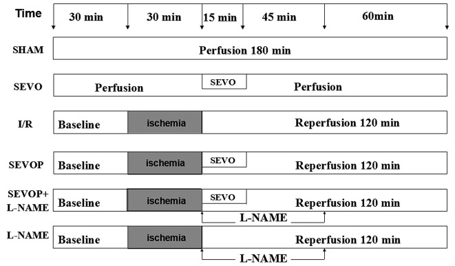
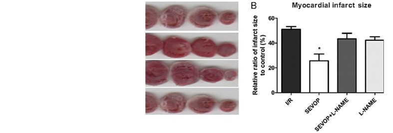
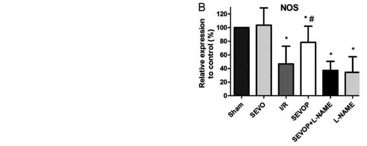
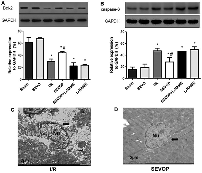
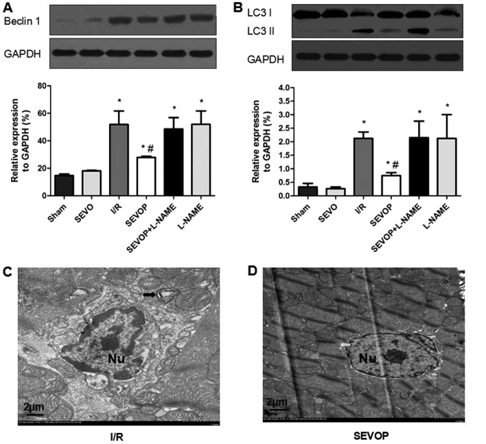
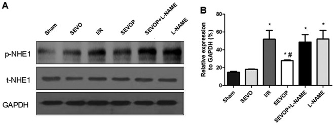
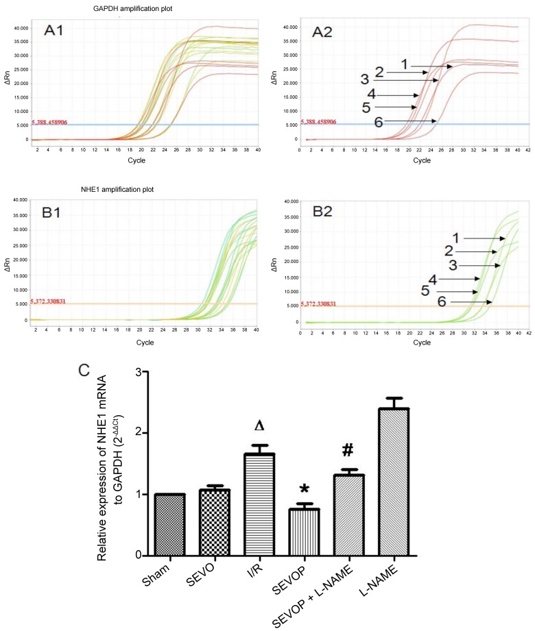
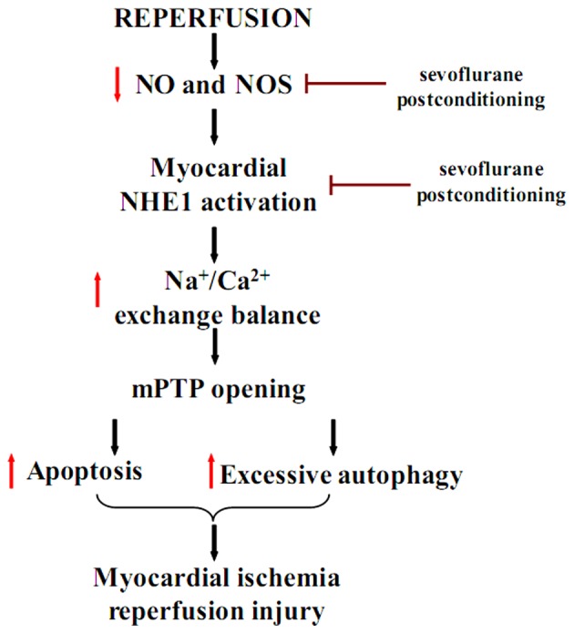
The protective effects of sevoflurane post-conditioning against myocardial ischemia/reperfusion (I/R) injury (MIRI) have been previously reported. However, the mechanisms responsible for these protective effects remain elusive. In this study, in order to investigate the molecular mechanisms responsible for the protective effects of sevoflurane post-conditioning on isolated rat hearts subjected to MIRI, Sprague-Dawley rat hearts were randomly divided into the following 6 groups: i) the sham-operated control; ii) 2.5% sevoflurane; iii) ischemia/reperfusion (I/R); iv) 2.5% sevoflurane post-conditioning plus I/R; v) 2.5% sevoflurane post-conditioning + NG-nitro-L-arginine methyl ester (L-NAME) plus I/R; and vi) L-NAME plus I/R. The infarct size was measured using 2,3,5-triphenyl tetrazolium chloride (TTC) staining. Additionally, the myocardial nitric oxide (NO), NO synthase (NOS) and nicotinamide adenine dinucleotide (NAD+) levels were determined. Autophagosomes and apoptosomes in the myocardium were detected by transmission electron microscopy. The levels of Bcl-2, cleaved caspase-3, Beclin-1, microtubule-associated protein light chain 3 (LC3)‑I/II, Na+/H+ exchanger 1 (NHE1) and phosphorylated NHE1 protein were measured by western blot analysis. NHE1 mRNA levels were measured by reverse transcription-quantitative polymerase chain reaction. Compared with the I/R group, 15 min of exposure to 2.5% sevoflurane during early reperfusion significantly decreased the myocardial infarct size, the autophagic vacuole numbers, the NHE1 mRNA and protein expression of cleaved caspase-3, Beclin-1 and LC3-I/II. Post-conditioning with 2.5% sevoflurane also increased the NO and NOS levels and Bcl-2 protein expression (p<0.05 or p<0.01). Notably, the cardioprotective effects of sevoflurane were partly abolished by the NOS inhibitor, L-NAME. The findings of the present study suggest that sevoflurane post-conditioning protects the myocardium against I/R injury and reduces the myocardial infarct size. The underlying protective mechanisms are associated with the inhibition of mitochondrial permeability transition pore opening, and with the attenuation of cardiomyoctye apoptosis and excessive autophagy. These effects are mediated through an increase in NOS and a decrease in phopshorylated NHE1 levels.


Figures

Figure 1
Figure 1
Experimental protocol. After randomly dividing the rats into 6 groups (n=24), rats underwent a 30-min ischemia procedure, followed by 120 min of reperfusion. The sham-operated group underwent a procedure without ischemia, but it was time-matched. Sevoflurane (2.5%) was inhaled for 15 min immediately following ischemia. Sham, sham-operated group; SEVO, sevoflurane group (no I/R injury); I/R, ischemia/reperfusion injury group; SEVOP, sevoflurane post-conditioning group; L-NAME, group treated with NG-nitro-L-arginine methyl ester (NOS inhibitor).
Figure 2
Figure 2
Sevoflurane post-conditioning decreases infarct size which is increased by myocardial ischemia/reperfusion injury (MIRI) in rats. (A) Representative TTC-stained images and (B) quantification of the percentage of the injured area. The columns and error bars represent the means ± SD; n=6 experiments. A one-way ANOVA followed by Tukey's post-hoc test was used to determine statistical significance. *P<0.05 vs. I/R. Sham, sham-operated group; SEVO, sevoflurane group (no I/R injury); I/R, ischemia/reperfusion injury group; SEVOP, sevoflurane post-conditioning group; L-NAME, group treated with NG-nitro-L-arginine methyl ester (NOS inhibitor); TTC, triphenyltetrazolium chloride.
Figure 3
Figure 3
Sevoflurane post-conditioning increases nitric oxide (NO) and nitric oxide synthase (NOS) production following ischemia/reperfusion. (A) Sevoflurane post-conditioning increased NO production. (B) Sevoflurane post-conditioning increased NOS production. Sevoflurane post-conditioning-induced NO and NOS production was inhibited by L-NAME treatment. Data are presented as the means ± SD; n=6 experiments. A one-way ANOVA followed by Tukey's post-hoc test was used to determine statistical significance. *P<0.05 vs. sham. #P<0.05 vs. I/R. Sham, sham-operated group; SEVO, sevoflurane group (no I/R injury); I/R, ischemia/reperfusion injury group; SEVOP, sevoflurane post-conditioning group; L-NAME, group treated with NG-nitro-L-arginine methyl ester (NOS inhibitor).
Figure 4
Figure 4
Alterations in Bcl-2 expression and cleaved-caspase-3 activity following sevoflurane post-conditioning. Shown are myocardial western blots of Bcl-2 and cleaved caspase-3 expression. (A and B) Representative western blots of Bcl-2 and cleaved-caspase-3, respectively, in Sham, SEVO, I/R, SEVOP, SEVOP+L-NAME and L-NAME groups. (C and D) Representative transmission electron microscopy images from the I/R and SEVOP groups, respectively (original magnification, ×2000; scale bar, 2 µm). Images show that apoptosomes were decreased in the SEVOP group when compared with the I/R group. As indicated by the arrows, panel (C) shows mild swelling of the nucleus, chromatin edge set along the nuclear membrane, loose cytoplasm and moderate swelling of the mitochondria These effects were attenuated in the SEVOP group (D). The data are presented as the means ± SD (n=5 experiments). A one-way ANOVA followed by Tukey's post-hoc test was used to determine statistical significance. *P<0.05 vs. sham. #P<0.05 vs. I/R. SEVO, sevoflurane group (no I/R injury); I/R, ischemia/reperfusion injury group; SEVOP, sevoflurane post-conditioning group; L-NAME, group treated with NG-nitro-L-arginine methyl ester (NOS inhibitor); Nu, nucleus.
Figure 5
Figure 5
Alterations in LC3-II/I and Beclin-1 expression following sevoflurane post-conditioning. Shown are myocardial western blots of LC3-II/I and Beclin-1 expression. (A and B) Representative western blots of Beclin-1 and LC3II/I, respectively, in the Sham, SEVO, I/R, SEVOP, SEVOP + L-NAME and L-NAME groups. Values are expressed as the means ± SD (n=5 experiments). A one-way ANOVA followed by Tukey's post-hoc test was used to determine statistical significance. *P<0.05 vs Sham. #P<0.05 vs I/R. (C and D) Representative transmission electron microscopy images from the I/R and SEVOP groups, respectively (original magnification, ×2,000; scale bar, 2 µm), showing that the number of autophagic vacuoles (AVs) decreased in the sevoflurane post-conditioning group when compared with the I/R group. (C) Arrow indicates autophagic vacuole. Sham, sham-operated group; SEVO, sevoflurane group (no I/R injury); I/R, ischemia/reperfusion injury group; SEVOP, sevoflurane post-conditioning group; L-NAME, group treated with NG-nitro-L-arginine methyl ester (NOS inhibitor); Nu, nucleus.
Figure 6
Figure 6
Sevoflurane post-conditioning inhibits p-NHE1 expression. (A and B) Shown are myocardial western blots of phosphorylated (p-)and total (t-)NHE1 expression. Values are expressed as the means ± SD. A one-way ANOVA followed by Tukey's post-hoc test was used to determine statistical significance. *P<0.05 vs. Sham. #P<0.05 vs. I/R. Sham, sham-operated group; SEVO, sevoflurane group (no I/R injury); I/R, ischemia/reperfusion injury group; SEVOP, sevoflurane post-conditioning group; L-NAME, group treated with NG-nitro-L-arginine methyl ester (NOS inhibitor).
Figure 7
Figure 7
Sevoflurane post-conditioning inhibits NHE1 mRNA expression. (A1) Representative amplification plot of GAPDH; 3 microwells/group, 6 groups; 3 different colors indicate 3 microwells. (A2) Representative amplification plot of GAPDH for 6 groups. Numbers 1, 2, 3, 4, 5 and 6, represent the amplification plot of GAPDH for the Sham, SEVO, I/R, SEVOP, SEVOP + L-NAME and L-NAME groups, respectively. (B1) Representative amplification plot of NHE1; 3 microwells/ group, 6 groups; 3 different colors indicate 3 microwells. (B2) Representative amplification plot of NHE1 for 6 groups. Numbers 1, 2, 3, 4, 5 and 6, represent the amplification plot of NHE1 for the Sham, SEVO, I/R, SEVOP, SEVOP + L-NAME and L-NAME groups, respectively. (C) mRNA expression of NHE1 relative to GAPDH, in the Sham, SEVO, I/R, SEVOP, SEVOP + L-NAME and L-NAME groups, respectively. Values are expressed as the means ± SD (n=3). A one-way ANOVA followed by Tukey's post-hoc test was used to determine statistical significance. ΔP<0.05 vs. Sham, *P<0.05 vs. I/R, #P<0.05 vs. SEVOP..
Figure 8
Figure 8
Proposed mechanism of sevoflurane post-conditioning cardioprotection against myocardial ischemia/reperfusion injury (MIRI). Black arrows indicate transition; red downward arrows indicate downregulation; red upward arrows indicate upregulation; and red bar-headed lines indicate inhibition.

References

    1. Hu Q, Chen J, Jiang C, Liu HF. Effect of peroxisome proliferator-activated receptor gamma agonist on heart of rabbits with acute myocardial ischemia/reperfusion injury. Asian Pac J Trop Med. 2014;7:271–275. doi: 10.1016/S1995-7645(14)60036-5. - DOI - PubMed
    1. Zhu M, Feng J, Lucchinetti E, Fischer G, Xu L, Pedrazzini T, Schaub MC, Zaugg M. Ischemic postconditioning protects remodeled myocardium via the PI3K-PKB/Akt reperfusion injury salvage kinase pathway. Cardiovasc Res. 2006;72:152–162. doi: 10.1016/j.cardiores.2006.06.027. - DOI - PubMed
    1. Xia A, Xue Z, Wang W, Zhang T, Wei T, Sha X, Ding Y, Zhou W. Naloxone postconditioning alleviates rat myocardial ischemia reperfusion injury by inhibiting JNK activity. Korean J Physiol Pharmacol. 2014;18:67–72. doi: 10.4196/kjpp.2014.18.1.67. - DOI - PMC - PubMed
    1. Yao YY, Zhu MH, Zhang FJ, Wen CY, Ma LL, Wang WN, Wang CC, Liu XB, Yu LN, Qian LB, et al. Activation of Akt and cardioprotection against reperfusion injury are maximal with only five minutes of sevoflurane postconditioning in isolated rat hearts. J Zhejiang Univ Sci B. 2013;14:511–517. doi: 10.1631/jzus.B1200195. - DOI - PMC - PubMed
    1. Sakai EM, Connolly LA, Klauck JA. Inhalation anesthesiology and volatile liquid anesthetics: Focus on isoflurane, desflurane, and sevoflurane. Pharmacotherapy. 2005;25:1773–1788. doi: 10.1592/phco.2005.25.12.1773. - DOI - PubMed

Publication types

MeSH terms