

Evaluation of prognostic factors and survival rates in malignant feline mammary gland neoplasms
- PMID: 26460079
- PMCID: PMC11112230
- DOI: 10.1177/1098612X15610367
Evaluation of prognostic factors and survival rates in malignant feline mammary gland neoplasms
Abstract
Objectives: The aim of the study was to investigate prognostic factors in feline mammary gland neoplasms, correlating them with overall survival (OS).
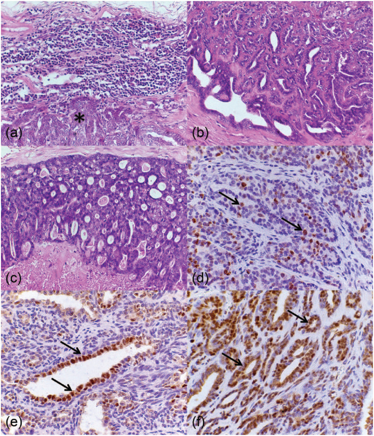
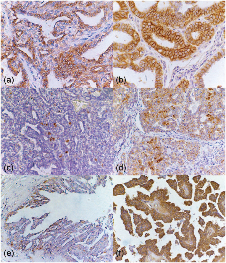
Methods: Fifty-six primary malignant mammary gland neoplasms and 16 metastatic lymph nodes from 37 female cats were analyzed. Clinical staging, histologic type and grade, and immunohistochemistry for Ki-67, progesterone and estrogen receptor, human epidermal growth factor receptor type 2 (HER-2), cyclooxygenase-2 (COX-2) and vascular endothelial growth factor (VEGF) were evaluated. Follow-up was performed in order to correlate prognostic factors with OS.
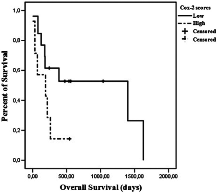
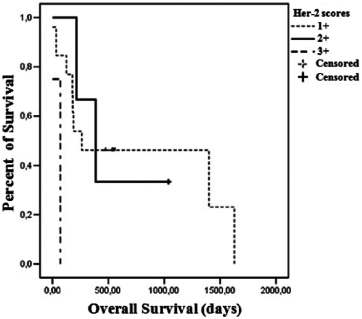
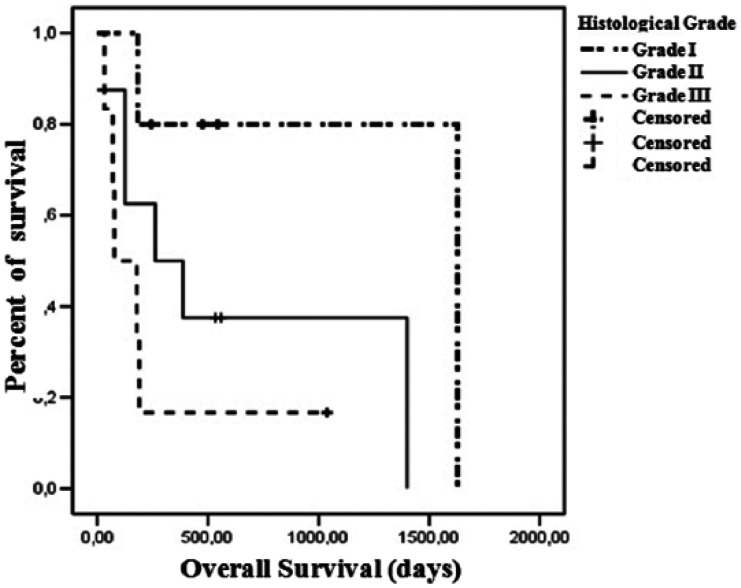
Results: Lymph node metastasis was found in 35% of cases. Clinical stage III, tubulopapillary carcinomas and histologic grade II cases prevailed in the study. Most neoplasms were positive for hormonal receptors, negative for HER-2 overexpression and presented VEGF overexpression. Immunoreactivity for Ki-67 (P = 0.046) and COX-2 (P = 0.007) was higher in metastases than in primary tumors. COX-2 (P = 0.089), HER-2 (P = 0.012) and histologic grade (P = 0.080) were correlated with OS.
Conclusions and relevance: The data suggest that inhibition of ovarian hormones and COX-2 may represent a therapeutic option for malignant feline mammary gland neoplasms. When evaluating disease progression, COX-2 scores and Ki-67 index should be analyzed in primary tumors and metastases. Histologic grade, HER-2 status and COX-2 scores were found to have a direct influence on OS. Prognostic factors allow for a better understanding of disease outcome in a condition that is characterized by a poor prognosis. The present work highlights the need for further studies on endocrine therapy and COX-2 inhibitors, which could influence OS.
© The Author(s) 2015.
Conflict of interest statement
The authors declared no potential conflicts of interest with respect to the research, authorship, and/or publication of this article.
Figures
Similar articles
-
Expression of HIF-1α and VEGF in feline mammary gland carcinomas: association with pathological characteristics and clinical outcomes.BMC Vet Res. 2020 May 6;16(1):125. doi: 10.1186/s12917-020-02338-y. BMC Vet Res. 2020. PMID: 32375802 Free PMC article.
-
COX-2 expression in canine and feline invasive mammary carcinomas: correlation with clinicopathological features and prognostic molecular markers.Breast Cancer Res Treat. 2006 Jul;98(1):115-20. doi: 10.1007/s10549-005-9138-z. Epub 2006 Mar 15. Breast Cancer Res Treat. 2006. PMID: 16538539
-
Grade is an independent prognostic factor for feline mammary carcinomas: a clinicopathological and survival analysis.Vet J. 2011 Jan;187(1):65-71. doi: 10.1016/j.tvjl.2009.10.030. Epub 2009 Dec 1. Vet J. 2011. PMID: 19955006
-
Prognostic evaluation of feline mammary carcinomas: a review of the literature.Vet Pathol. 2015 Jan;52(1):46-60. doi: 10.1177/0300985814528221. Epub 2014 Apr 16. Vet Pathol. 2015. PMID: 24741029 Review.
-
Prognostic histopathological and molecular markers in feline mammary neoplasia.Vet J. 2012 Oct;194(1):19-26. doi: 10.1016/j.tvjl.2012.05.008. Epub 2012 Jul 28. Vet J. 2012. PMID: 22841451 Review.
Cited by
-
Regional Variations in and Key Predictors of Feline Tumor Malignancy: A Decade-Long Retrospective Study in Korea.Animals (Basel). 2024 Oct 16;14(20):2989. doi: 10.3390/ani14202989. Animals (Basel). 2024. PMID: 39457919 Free PMC article.
-
The role of COX expression in the prognostication of overall survival of canine and feline cancer: A systematic review.Vet Med Sci. 2021 Jul;7(4):1107-1119. doi: 10.1002/vms3.460. Epub 2021 Mar 10. Vet Med Sci. 2021. PMID: 33751829 Free PMC article.
-
Sentinel Lymph Node Mapping and Biopsy in Cats with Solid Malignancies: An Explorative Study.Animals (Basel). 2022 Nov 11;12(22):3116. doi: 10.3390/ani12223116. Animals (Basel). 2022. PMID: 36428344 Free PMC article.
-
Expression of HIF-1α and VEGF in feline mammary gland carcinomas: association with pathological characteristics and clinical outcomes.BMC Vet Res. 2020 May 6;16(1):125. doi: 10.1186/s12917-020-02338-y. BMC Vet Res. 2020. PMID: 32375802 Free PMC article.
-
Canine, Feline, and Murine Mammary Tumors as a Model for Translational Research in Breast Cancer.Vet Sci. 2025 Feb 19;12(2):189. doi: 10.3390/vetsci12020189. Vet Sci. 2025. PMID: 40005948 Free PMC article. Review.
References
-
- Macewen EG, Hayes AA, Harvey HJ, et al.. Prognostic factors for feline mammary tumours. J Am Vet Med Assoc 1984; 185: 201–204. - PubMed
-
- Misdorp W. Tumors of the mammary gland. In: Meuten DJ. (ed). Tumors in domestic animals. 4th ed. Ames, IA: Iowa State Press, 2002, pp 575–606.
-
- Overley B, Shofer FS, Goldschmidt MH, et al.. Association between ovariohysterectomy and feline mammary carcinoma. J Vet Intern Med 2005; 19: 560–563. - PubMed
-
- Lana SE, Rutteman GR, Withrow SJ. Tumors of the mamary gland. In: Withrow FJ, Vail DM. (eds). Withrow & MacEwen’s small animal clinical oncology. 4th ed. Philadelphia, PA: WB Saunders, 2007, pp 619–636.
-
- Bostock DE. Canine and feline mammary neoplasms. Br Vet J 1986; 142: 506–515. - PubMed
Publication types
MeSH terms
Substances
LinkOut - more resources
Full Text Sources
Other Literature Sources
Research Materials
Miscellaneous
