. 2015 Oct 12;28(4):529-540.
doi: 10.1016/j.ccell.2015.09.006.

Comprehensive Genomic Characterization of Long Non-coding RNAs across Human Cancers

Affiliations

Comprehensive Genomic Characterization of Long Non-coding RNAs across Human Cancers

Xiaohui Yan et al. Cancer Cell. .

Abstract

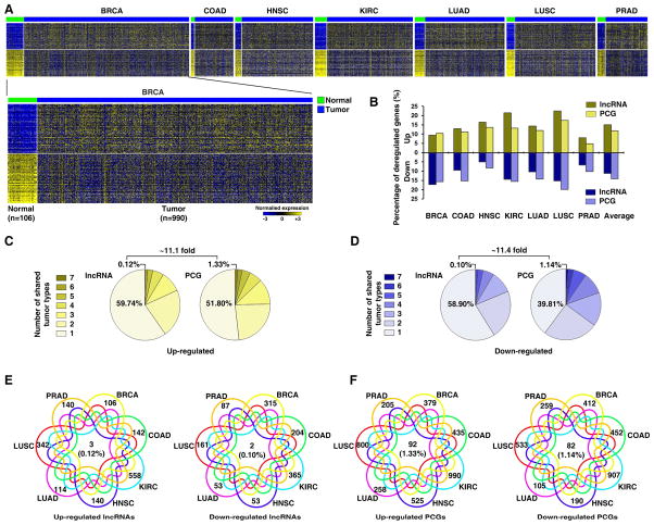
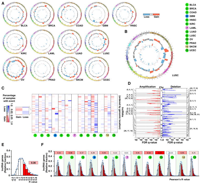
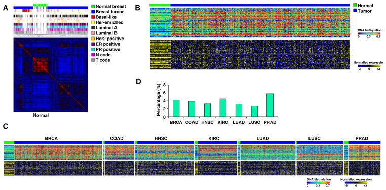
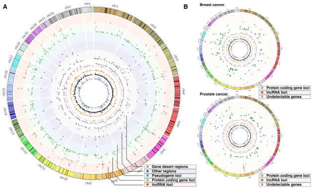
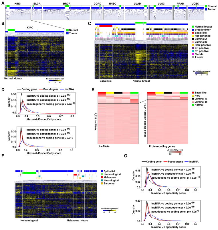
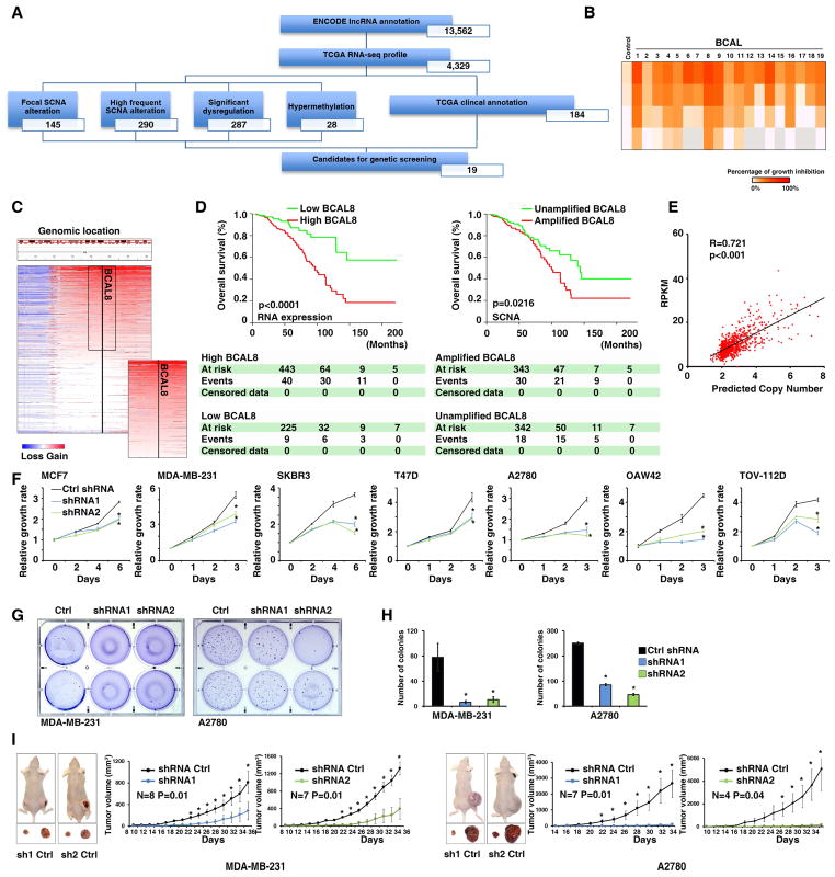
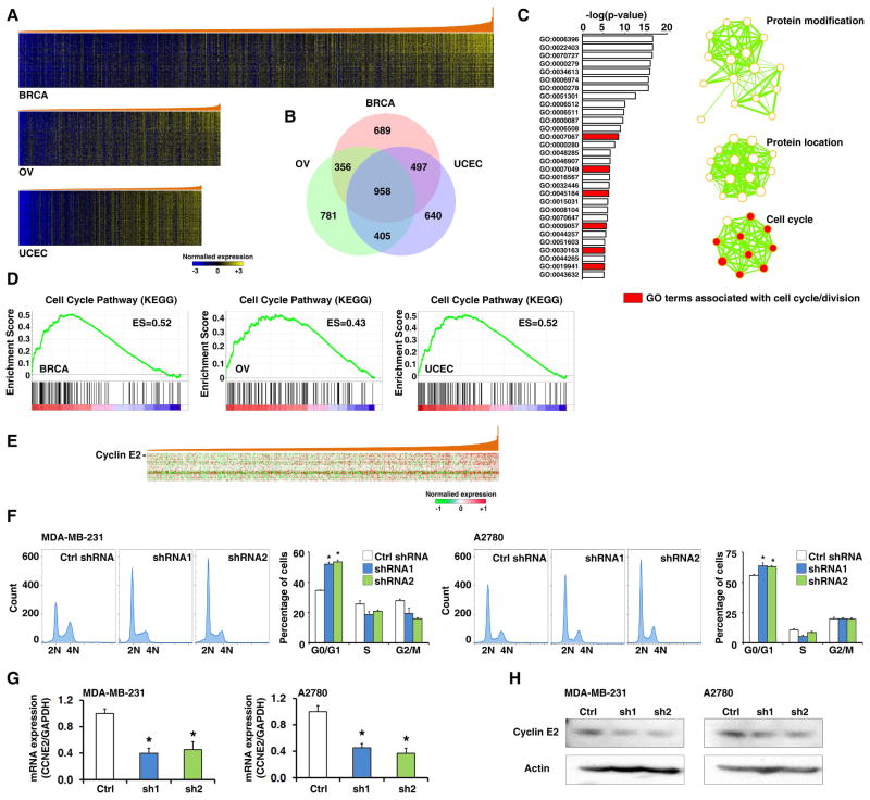
The discovery of long non-coding RNA (lncRNA) has dramatically altered our understanding of cancer. Here, we describe a comprehensive analysis of lncRNA alterations at transcriptional, genomic, and epigenetic levels in 5,037 human tumor specimens across 13 cancer types from The Cancer Genome Atlas. Our results suggest that the expression and dysregulation of lncRNAs are highly cancer type specific compared with protein-coding genes. Using the integrative data generated by this analysis, we present a clinically guided small interfering RNA screening strategy and a co-expression analysis approach to identify cancer driver lncRNAs and predict their functions. This provides a resource for investigating lncRNAs in cancer and lays the groundwork for the development of new diagnostics and treatments.


Figures

Figure 1
Figure 1. The expression of lncRNAs is dysregulated in cancer
A. Heatmap of lncRNAs whose expression is significantly dysregulated. The top 100 most significantly dysregulated lncRNAs from each individual tumor-type are presented. B. The percentages of the dysregulated lncRNAs and PCGs. C. and D. The percentages of the up- (C) and down- (D) regulated lncRNAs (left) and PCGs (right) that were shared among the seven cancer types. E. and F. Venn diagrams of the up- (left) and down- (right) regulated lncRNAs (E) and PCGs (F) shared among the seven cancer types. See also Figure S1 and Tables S1 and S2.
Figure 2
Figure 2. Somatic copy numbers of lncRNA genes are altered in cancer with different frequencies
A. A genome-wide view of SCNAs in cancers. The outer track shows the frequencies of SCNAs from the lncRNA-containing loci and the inner track shows the focal alteration regions. B. An enlarged view of SCNAs in LUSC. C. Heatmap of somatic copy number gain and loss for lncRNA genes. The rows, each of which represents an lncRNA gene locus, are arranged according to the genomic locations of the lncRNA genes. Left: frequency of gain (red); right: frequency of loss (blue). D. The lncRNA and PCGs in the top 20 focal gain (left) or loss (right) peaks in LUSC. The numbers of PCGs (left), annotated lncRNAs (middle), and detectable lncRNAs (right) in each peak are indicated in parentheses. E. and F. Histogram of percentage of lncRNAs whose RNA-SCNA correlation coefficients are in specific ranges across 13 cancer types (E) and in each cancer type (F). The number and red color intensity in the inserts indicate the percentage of the detectable lncRNAs whose Pearson’s R value was ≥0.2 in a given cancer type. See also Figure S2 and Tables S3.
Figure 3
Figure 3. DNA methylation patterns in the promoter regions of lncRNA genes are altered in cancer
A. NMF clustering of DNA methylation probes that are located in lncRNA promoters and whose methylation β-values had the largest variations across all breast specimens. B. Heatmaps of the methylation status (β-value, upper) in the promoter regions and the RNA expression level (lower) of the corresponding lncRNAs in breast specimens. C. Heatmaps of the methylation status of the lncRNA promoter regions and the RNA expression levels. D. A summary of the percentage of the CAESLG. See also Figure S3 and Tables S4 and S5.
Figure 4
Figure 4. Many cancer-associated SNPs are located in lncRNA loci
A. A genome-wide view of the most significant cancer-associated index SNPs. The peaks in each track are proportional to the p-values between the chromosomal locations of the index-SNPs. B. Genome-wide view of the breast (upper) and prostate (lower) index-SNPs in lncRNA (red) and PCG loci (green).
Figure 5
Figure 5. The expression of lncRNAs is a specific biomarker in cancer
A. Unsupervised hierarchical cluster analyses on the expression of the top 10% lncRNAs whose expression levels varied the most across all samples within each cancer type. B. Heatmap generated by unsupervised cluster analysis of lncRNAs with the largest expression variation in kidney cancer. C. Heatmap of unsupervised hierarchical cluster analysis using lncRNA signatures from breast cancer. D. Distribution of maximal subtype specificity scores calculated for each gene across the breast cancer specimens for all expressing transcripts (upper) or high expressers (lower) for lncRNA (blue), pseudogenes (red), and PCGs (black). E. Heatmap of lncRNA (left) and PCG (right) expression (JC scores) sorted on the basis of tissue-specific expression. Top: tissue-specific; bottom: ubiquitously expressed. F. Heatmap of unsupervised hierarchical cluster analysis using lncRNA signatures from the CCLE RNA-seq dataset. G. Distributions of maximal cancer-type specificity scores calculated for each gene across the CCLE major cancer types and across all expressing genes (upper) or high expressers (lower) for lncRNAs (blue), pseudogenes (red) and PCGs (black). See also Figure S4 and Tables S6.
Figure 6
Figure 6. An effective strategy to integrate multidisciplinary information from TCGA to identify cancer driver lncRNAs
A. Flowchart describing the process of candidate gene selection in breast cancer. B. The summary of the proof-of-concept siRNA screening in MDA-MB-231 cells. C. Copy number profiles of BCAL8 locus from breast tumor specimens. D. Survival curves of breast cancer patients with high and low BCAL8 RNA expression (left) and differing genomic SCNA status (right). The numbers of patients who were alive (at risk), deceased (event), or censored during the course of surveillance are indicated in the table under the corresponding time points. E. The correlation between BCAL8 gene copy number and RNA expression in breast cancer. F. The growth curves of cells expressing control or BCAL8 shRNAs. G. Soft-agar assays (in 6-well plates) on cells expressing control or BCAL8 shRNAs. H. Quantification of the number of colonies from the softer agar assays. I. Xenograft tumor growth of cells expressing control or BCAL8 shRNAs. Error bars: SD. *: p<0.05. See also Figure S5 and Tables S7.
Figure 7
Figure 7. Inferring the functions of BCAL8 by integrative bioinformatics analyses
A. Heatmap of PCGs that were significantly and positively co-expressed with BCAL8. The genes were arranged from top to bottom in ascending order of their correlation with BCAL8. B. Venn diagrams of BCAL8-associated genes among breast, ovarian, and endometrial cancers. C. Pathways over-represented by BCAL8-associated PCGs in all three cancer types according to DAVID analysis based on gene ontology term. D. Enrichment of cell cycle pathway genes in cancer specimens with high levels of BCAL8. E. Heatmap of PCGs whose protein expression (RPPA) is significantly correlated with BCAL8 expression in breast cancer. The proteins are arranged from top to bottom in ascending order of their correlation with BCAL8 expression. F. Cell-cycle profiles of cells expressing control and BCAL8 shRNAs. G. qRT-PCR of CCNE2 mRNA expression in cells expressing control or BCAL8 shRNAs. H. Western blot of Cylin E2 in cells expressing control or BCAL8 shRNAs. Error bars: SD. *: p<0.05. See also Figure S6 and Tables S8.

References

    1. Akrami R, Jacobsen A, Hoell J, Schultz N, Sander C, Larsson E. Comprehensive analysis of long non-coding RNAs in ovarian cancer reveals global patterns and targeted DNA amplification. PloS one. 2013;8:e80306. - PMC - PubMed
    1. Batista PJ, Chang HY. Long noncoding RNAs: cellular address codes in development and disease. Cell. 2013;152:1298–1307. - PMC - PubMed
    1. Cabili MN, Trapnell C, Goff L, Koziol M, Tazon-Vega B, Regev A, Rinn JL. Integrative annotation of human large intergenic noncoding RNAs reveals global properties and specific subclasses. Genes & development. 2011;25:1915–1927. - PMC - PubMed
    1. Calin GA, Liu CG, Ferracin M, Hyslop T, Spizzo R, Sevignani C, Fabbri M, Cimmino A, Lee EJ, Wojcik SE, et al. Ultraconserved regions encoding ncRNAs are altered in human leukemias and carcinomas. Cancer cell. 2007;12:215–229. - PubMed
    1. Derrien T, Johnson R, Bussotti G, Tanzer A, Djebali S, Tilgner H, Guernec G, Martin D, Merkel A, Knowles DG, et al. The GENCODE v7 catalog of human long noncoding RNAs: analysis of their gene structure, evolution, and expression. Genome research. 2012;22:1775–1789. - PMC - PubMed

Publication types

Substances