Review
. 2015 Oct;29(5):825-38.
doi: 10.1016/j.hoc.2015.06.003. Epub 2015 Aug 20.

Cell(s) of Origin of Langerhans Cell Histiocytosis

Affiliations
Review

Cell(s) of Origin of Langerhans Cell Histiocytosis

Matthew Collin et al. Hematol Oncol Clin North Am. 2015 Oct.

Abstract

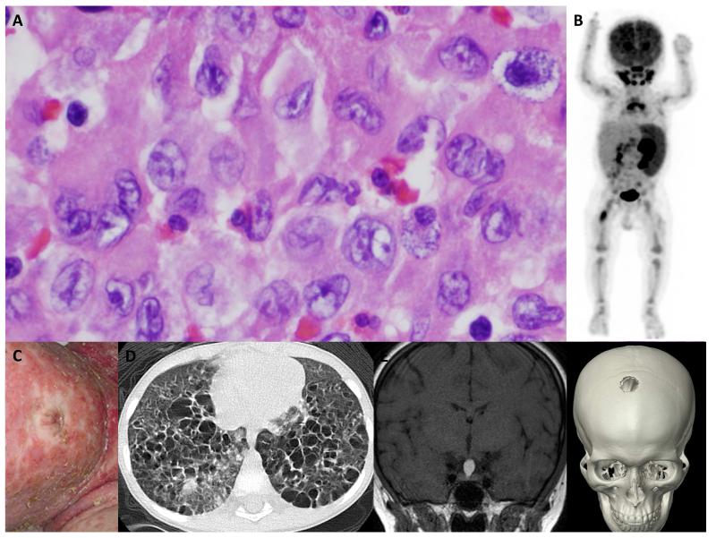
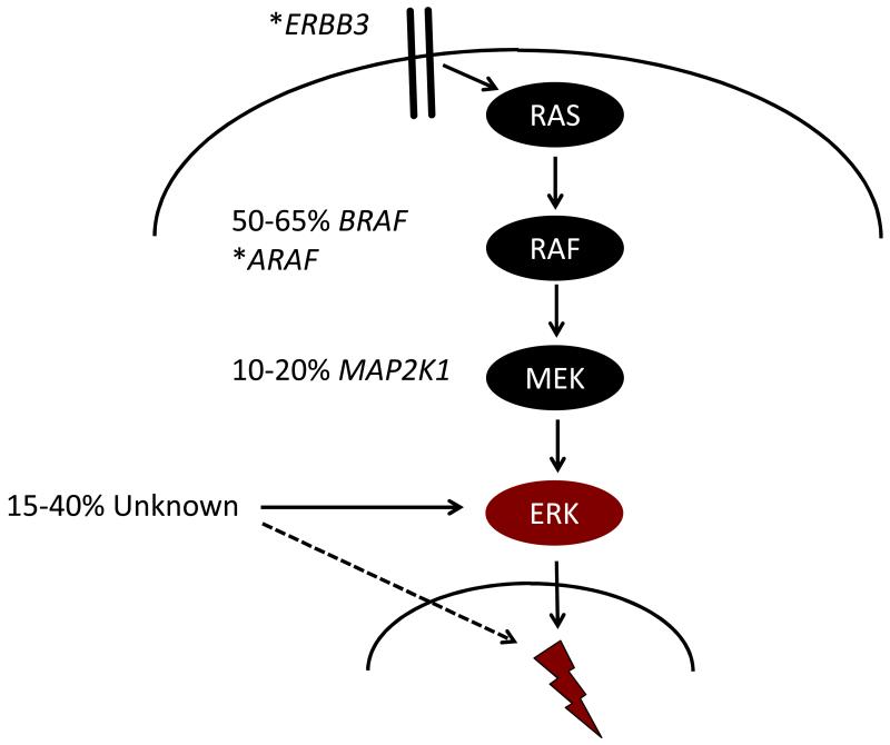
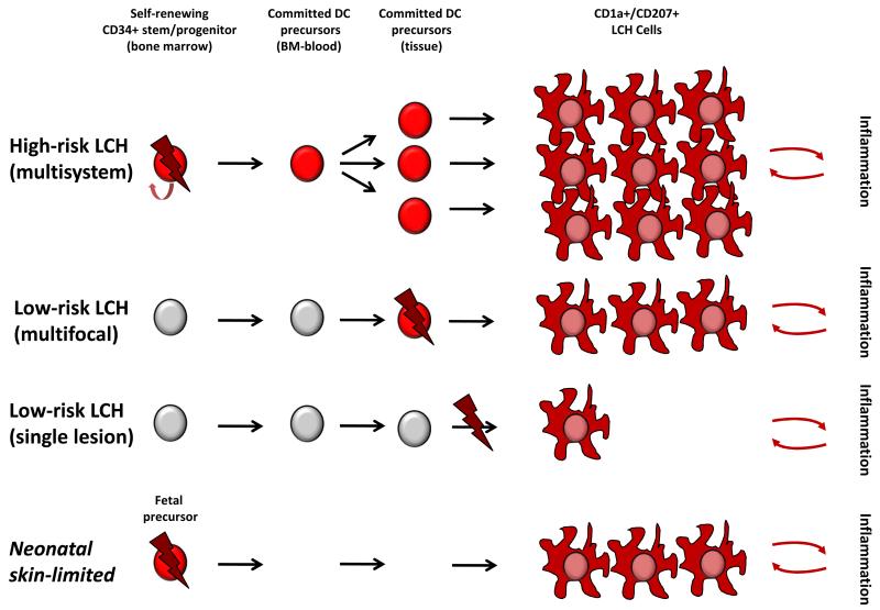
Langerhans cell histiocytosis (LCH) is heterogeneous disease characterized by common histology of inflammatory lesions containing Langerin(+) (CD207) histiocytes. Emerging data support a model in which MAPK activation in self-renewing hematopoietic progenitors may drive disseminated high-risk disease, whereas MAPK activation in more differentiated committed myeloid populations may induce low-risk LCH. The heterogeneous clinical manifestations with shared histology may represent the final common pathway of an acquired defect of differentiation, initiated at more than one point. Implications of this model include re-definition of LCH as a myeloid neoplasia and re-focusing therapeutic strategies on the cells and lineages of origin.

Keywords: BRAF; Dendritic cell; Langerhans cell histiocytosis; MAPK signaling; Myeloid differentiation.


Figures

Figure 1
Figure 1. LCH: Common histology and clinical heterogeneity
A. Hematoxylin and eosin staining of a typical LCH lesion, demonstrating the classic large histiocytes with grooved “coffee-bean” nuclei and abundant eosinophilic cytoplasm. The inflammatory infiltrate varies, but typically includes lymphocytes, eosinophils and macrophages. Histologic features have not been associated with specific clinical presentations. This biopsy could have come from any of the cases presented in B-F. B. PET scan of a 1 year old with high-risk LCH: PET-avid R femur lesion, spleen, bone marrow and cervical lymph nodes are evident in this image. C. Infant with severe LCH skin lesions. In infants, skin LCH may be self-limiting and spontaneously resolve or may be part of life-threatening multi-system high-risk disease. D. CT scan demonstrating innumerable cysts and lung lesions in a 3 year old with LCH involving lung, pituitary and skin. E. Brain MRI in a teenager with an isolated pituitary LCH lesion. F. CT scan of a teenager with an isolated skull LCH lesion.
Figure 2
Figure 2. Model of LCH pathogenesis: ERK activation at specific stages of myelopoiesis determines clinical phenotype
A. Mutually exclusive activating mutations in MAPK genes have been reported in most cases of LCH, and ERK activation appears to be universal in LCH lesion CD207+ cells. Lightning bolt represents the downstream impact of ERK activation that presumably is essential for LCH pathogenesis. B. Proposed “Misguided Myeloid DC” model of LCH pathogenesis in which somatic mutation (lightning bolt) and subsequent ERK activation (red cell) at specific stages of DC development determines clinical outcome. This model reflects interpretation of data from human and mouse studies, but leaves room for future refinement. According to this model, ERK activation in a self-renewing progenitor/stem cell in the bone marrow has the potential to form lesions in hematopoietic organs, liver, and virtually any organ system leading to MS LCH with somatic mutation detectable in BM and blood. In contrast, ERK activation in committed precursors may form multiple lesions in a limited number of organ systems, but somatic mutation is not usually detectable in the BM or blood at diagnosis, or subsequently. ERK activation at a later stages of differentiation, perhaps even in a single tissue cell, may form a single unifocal lesion. Based on recent data describing the pre-natal origin of tissue myeloid cells, it is also conceivable that ERK activating mutation may arise during fetal development. This is speculative but could explain “self-resolving” neonatal LCH (Hashimoto Pritzker syndrome) in which mutated fetal precursors would be replaced by normal myeloid cells after birth. In all models, it is suggested that LCH cells recruit and activate inflammatory cells which may provide reciprocal survival signals and clearly play a role in clinical manifestations of LCH. Data from Refs -,

Similar articles

Cited by

References

    1. Van Furth R, Cohn ZA. The origin and kinetics of mononuclear phagocytes. J.Exp.Med. 1968;128:415–435. - PMC - PubMed
    1. Guilliams M, Ginhoux F, Jakubzick C, et al. Dendritic cells, monocytes and macrophages: a unified nomenclature based on ontogeny. Nat.Rev.Immunol. 2014;14:571–578. - PMC - PubMed
    1. Geissmann F, Manz MG, Jung S, et al. Development of monocytes, macrophages, and dendritic cells. Science. 2010;327:656–661. - PMC - PubMed
    1. Collin M, McGovern N, Haniffa M. Human dendritic cell subsets. Immunology. 2013;140:22–30. - PMC - PubMed
    1. Nezelof C, Basset F, Rousseau MF. Histiocytosis × histogenetic arguments for a Langerhans cell origin. Biomedicine. 1973;18:365–371. - PubMed

Publication types